BrightER - Endoplasmic reticulum probe for live imaging & flow cytometry
Reticulum endoplasmic probe for live imaging and flow cytometry
BrightER is a selective, cell-permeable endoplasmic reticulum probe conjugated to a bright photostable rhodamine dye. Simply add it to your culture medium and get a bright, stable ER fluorescence for at least 3 hours without altering ER physiology.
BrightER is suitable for both live imaging & flow cytometry experiments.
Technical information
Colour: Red
Detection method: Fluorescent
Wavelength range: Ex:557/Em:576.
Dye type: Tetramethylrhodamine
Assays: confocal imaging, flow cytometry
Form: Liquid
SubCellular localization: Endoplasmic reticulum
Lifetime: up to 4 months at -20°C
Kit content:
One vial of 100µL BrightER 5mM in DMSO+PEG solution.
Fixable: no
One BrightER kit is sufficient to prepare 10 mL of final imaging medium when used at the recommended 50 µM concentration.
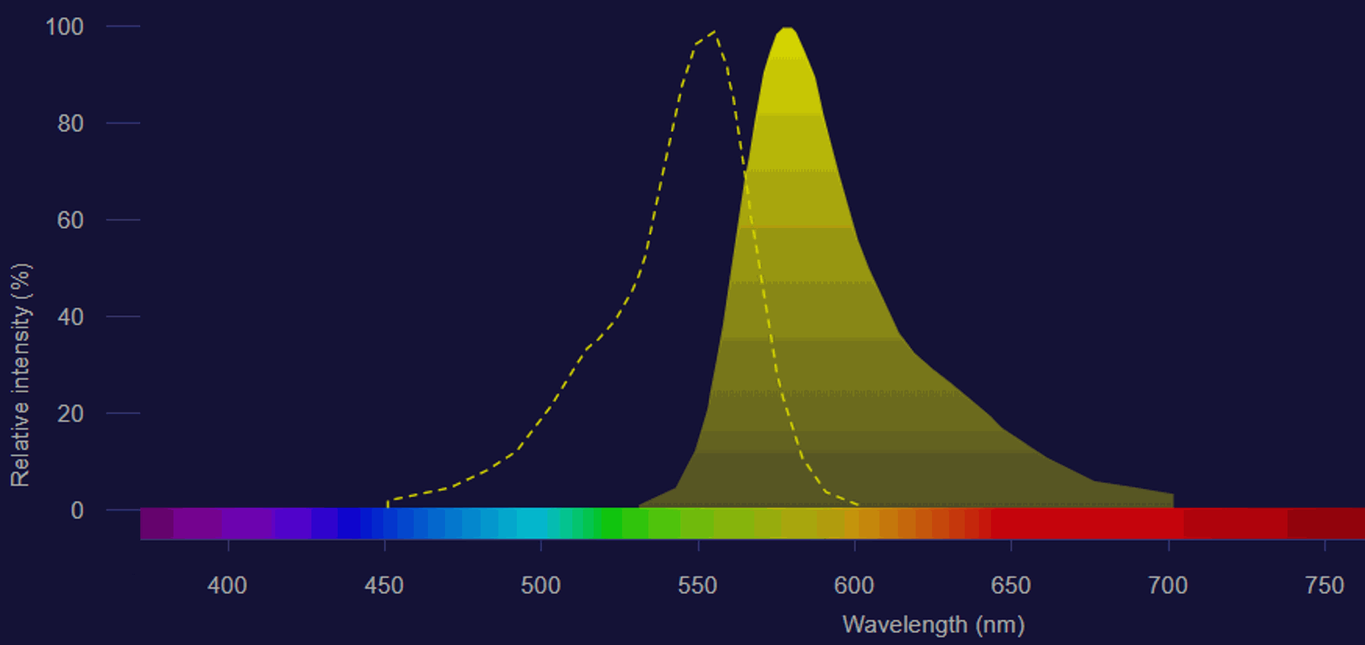
Absorption (dotted line) and emission (solid line) spectrum of BrightER
Image obtained on Fluorescence SpectraViewer (Thermo Fisher Scientific)
Applications
- Live imaging: ER morphological features and dynamic processes (membrane contact sites, intracellular trafficking, secretory pathways, viral infections, translation, protein folding and unfolded protein response (UPR), nuclear envelop formation upon mitosis, cellular calcium regulation and many others).
- Flow cytometry: quantification assays (ER stress detection, ER mass, co-localization studies)
Tested and validated on (cell types): Patient-derived glioblastoma cells, HEK293, HeLa, U-2 OS (human osteosarcoma), SVG-A (human astrocytes), SUM159 (human breast cancer cells), B16F10 (murine melanoma), LLC (Lewis lung carcinoma) & Vero E6 (monkey kidney epithelial cells).
Suggestions for use: The BrightER dye may be added directly in full media. In most cases, a final 50µM concentration is sufficient for immediate and bright ER staining; however, optimization may be needed for some cell types, conditions, and applications. BrightER is detected through standard TRITC and DsRed filters.
Additional resources:
> Safety Datasheet
BrightER staining in live HeLa cells (60X objective)
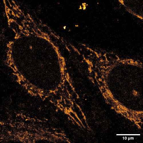
Image credit: Raphaël Gaudin & Yonis Bare – IRIM CNRS, Université de Montpellier - 2023
BrightER staining in live HeLa cells
HeLa cells were stained with BrightER (red), LysoTracker green (green) and imaged using a Zeiss LSM880 confocal laser scanning microscope in Airyscan mode (63X, 7 frames per second, interval 250ms)
Image credits: Céline Vrancx, PhD - Laboratory for Membrane Trafficking - Pr. Wim Annaert, VIB Center for Brain & Disease Research, KU Leuven Department of Neuroscience, Campus Gasthuisberg O&N5 Herestraat 49, 3000 Leuven, Belgium
BrightER staining in live SVG-A human astrocytes
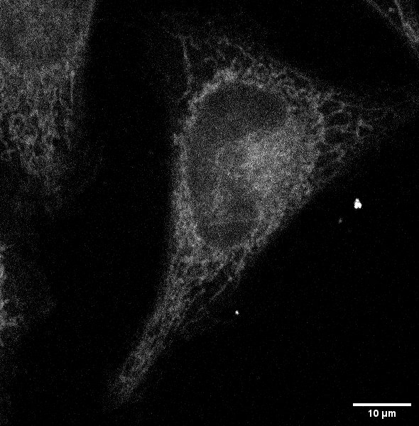
Image credit: Raphaël Gaudin & Yonis Bare – IRIM CNRS, Université de Montpellier - 2022
Assessing ER stress response after Thapsigargin treatment in mice cells

B16F10 (left) or LLC (right) cells were incubated with BrightER for 30min in HBSS after being treated with vehicle (DMSO) or Thapsigargin (1, 2 or 5µM) for 6h. Changes in mean fluorescence intensity (MFI) were assessed by flow cytometry using a CytoFLEX instrument.
Image credit: Jessica Mandula & Dr. Paulo Rodriguez - Moffitt Cancer Center, Florida (US) - 2023
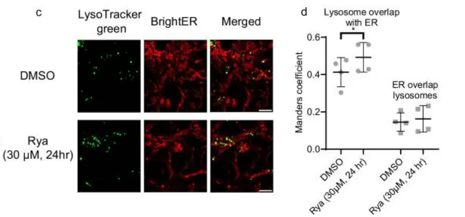
Figure 7c shows representative live‑cell Airyscan microscopy images of neurons stained with LysoTracker Green (lysosomes) and BrightER (endoplasmic reticulum), demonstrating how the ER and lysosomes spatially relate under control versus RyR‑inhibited conditions.
Figure 7d quantifies the colocalization (Manders overlap coefficients) between the BrightER ER signal and the LysoTracker lysosomal signal, showing that inhibiting RyR activity increases ER–lysosome contact site formation.
Credit: Inactive ryanodine receptors sustain lysosomal availability for autophagy by promoting ER-lysosomal contact site formation
Tim Vervliet et al.
Link: doi: 10.1038/s41467-025-68054-z
BrightER does not affect intracellular protein trafficking in breast cancer cells
![]()
A. The strong co-localisation of BrightER and Transferrin Receptor (TfR) fluorescence signals shows ER localisation of TfR, before biotin addition, in SUM159 cells CRISPR-edited to express the TfR-eRUSH system*. B. BrightER labeling does not alter biotin-induced TfR trafficking from the ER to the plasma membrane.
Image credit: Raphaël Gaudin & Yonis Bare – IRIM CNRS, Université de Montpellier - 2023
*link: https://www.science.org/doi/pdf/10.1126/sciadv.aba7803
BrightER staining in live Vero E6 monkey kidney epithelial cells
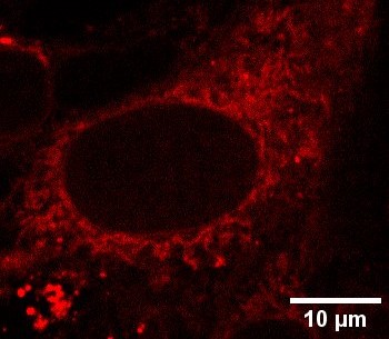
Image credit: Raphaël Gaudin & Yonis Bare – IRIM CNRS, Université de Montpellier - 2022
BrightER staining in live SVG-A human astrocytes
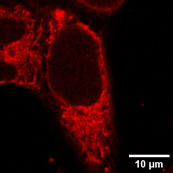
Image credit: Raphaël Gaudin & Yonis Bare – IRIM CNRS, Université de Montpellier - 2022
High-resolution microscopy picture showing BrightER (red) and commercial ER staining kit Cytopainter (green) signals in HeLa cells
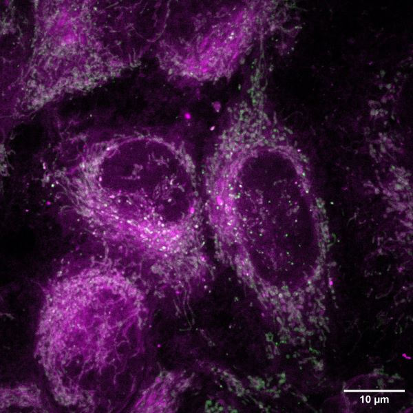
Image credit: Raphaël Gaudin & Yonis Bare – IRIM CNRS, Université de Montpellier - 2023
How to use BrightER in pictures
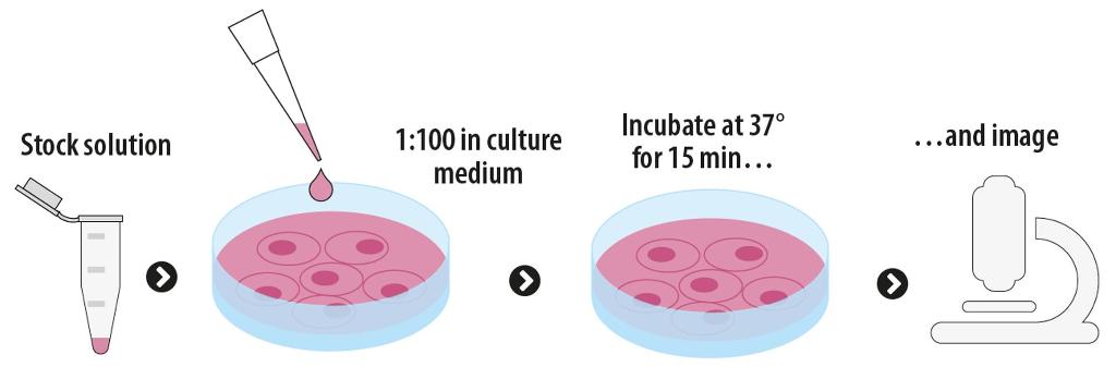
-> Read the full protocol: BrightER protocol
Everspark technology has been intially developed by Karine Monier, Arnaud Favier and Christophe Place and and published in Scientific Reports:
Provost, A., Rousset, C., Bourdon, L. et al. Innovative particle standards and long-lived imaging for 2D and 3D dSTORM. Sci Rep 9, 17967 (2019). https://doi.org/10.1038/s41598-019-53528-0
Publications:
Nanoscale engagement and clusterization of Programmed death ligand 1 (PD-L1) in the membrane lipid rafts of Non-Small Cell Lung Cancer cells
Martina Ruglioni, Simone Civita, Tiziano Salvadori, Sofia Cristiani, Vittoria Carnicelli, Serena Barachini, Iacopo Petrini, Irene Nepita, Marco Castello, Alberto Diaspro, Paolo Bianchini, Barbara Storti, Ranieri Bizzarri, Stefano Fogli and Romano Danesi bioRxiv 2022.08.09.503318; doi: https://doi.org/10.1101/2022.08.09.503318
HIV-1 diverts actin debranching mechanisms for particle assembly and release in CD4 T lymphocytes
Rayane Dibsy, Erwan Bremaud, Johnson Mak, Cyril Favard, Delphine Muriaux
BioRxiv, December 16, 2022. doi. 10.1101/2022.12.15.520580
Fluorescent Polymer-AS1411-Aptamer Probe for dSTORM Super-Resolution Imaging of Endogenous Nucleolin
Fabre L, Rousset C, Monier K, Da Cruz-Boisson F, Bouvet P, Charreyre MT, Delair T, Fleury E, Favier A. Biomacromolecules. 2022 May 12. doi: 10.1021/acs.biomac.1c01706. PMID: 35549176
Comparative analysis of ChAdOx1 nCoV-19 and Ad26.COV2.S SARS-CoV-2 vector vaccines.
Michalik S, Siegerist F, Palankar R, Franzke K, Schindler M, Reder A, Seifert U, Cammann C, Wesche J, Steil L, Hentschker C, Gesell-Salazar M, Reisinger E, Beer M, Endlich N, Greinacher A, Völker U. Haematologica. 2022 Apr 1;107(4):947-957. doi: 10.3324/haematol.2021.280154. PMID: 35045692
Metabolic biorthogonal labeling and dSTORM imaging of peptidoglycan synthesis in Streptococcus pneumoniae
Jennyfer Trouve, Oleksandr Glushonkov and Cecile Morlot
Star Protocols, December 13, 2021. doi: 10.1016/j.xpro.2021.101006
Insights in ChAdOx1 nCoV-19 vaccine-induced immune thrombotic thrombocytopenia.
Greinacher A, Selleng K, Palankar R, Wesche J, Handtke S, Wolff M, Aurich K, Lalk M, Methling K, Völker U, Hentschker C, Michalik S, Steil L, Reder A, Schönborn L, Beer M, Franzke K, Büttner A, Fehse B, Stavrou EX, Rangaswamy C, Mailer RK, Englert H, Frye M, Thiele T, Kochanek S, Krutzke L, Siegerist F, Endlich N, Warkentin TE, Renné T.
Blood. 2021 Dec 2;138(22):2256-2268. doi: 10.1182/blood.2021013231. PMID: 34587242
Superresolution Microscopy of Drosophila Indirect Flight Muscle Sarcomeres.
Szikora S, Novák T, Gajdos T, Erdélyi M, Mihály J. Bio Protoc. 2020 Jun 20;10(12):e3654. doi: 10.21769/BioProtoc.3654. eCollection 2020 Jun 20. PMID: 33659324