
Chitozen - Adhesive coverslips for bacteria imaging
Chitosan-coated coverslips for live bacteria imaging
For any other kit format request, please contact us
A technology designed by Tâm Mignot, Olivier Theodoly, Amandine Desorme, Guillaume Sudre and Laurent David.
Chitozen is a chitosan-coated coverslip immobilizing bacteria for microscopy without inducing bacteriostatic effects. It is compatible with 6-channel sticky slides for flow experiments. It is very helpful if you want to:

Increased Stability
Maintain bacteria in a same focal plane for imaging while preserving their physiology

Bacterial Imaging
Image bacteria both still and alive under the microscope

Renew culture medium and change growth condition
during the experiment and directly observe, in real-time, the bacteria new comportment under the microscope

Capture the moment
fix your bacteria and label them in situ to look at specific protein expression patterns during dynamic processes
Kit contents of Chitozen
Chitozen coverslips are compatible with 6-channel sticky slides to work in a closed system in static or dynamic flow conditions. Order you coverslips alone if you need to work in an open system (i.e. for AFM imaging) or to place them on depression slides.
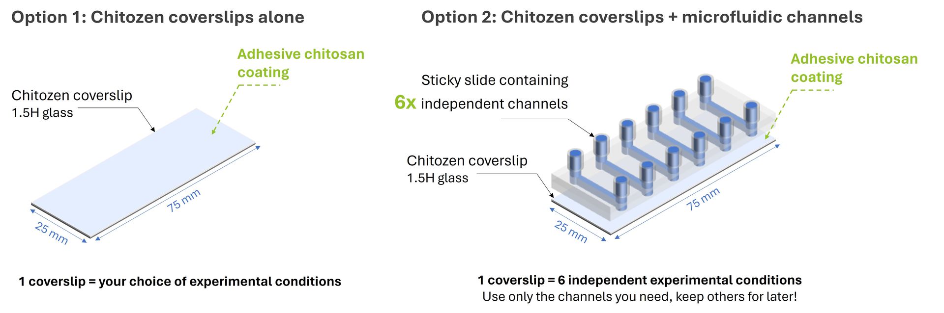
- 5-coverslips alone*
> 5x standard (25 x 75 mm) chitosan-coated coverslips - 5-coverslips with microfluidic channels (= 30 experiments)
> 5x standard (25 x 75 mm) chitosan-coated coverslips
> 5x bottomless 6-channel sticky slides
- (Optional) centrifugation pack: the Chitozen coverslips can be centrifuged using a rack compatible with standard microscope slide (25mmx75mm) dimensions. If needed, add our centrifugation pack to your Chitozen kit. It includes:
> µ-Slide Microscopy Rack (ibidi)
> Magnetic Lid for Microscopy Rack
> Clamp & adapter to securely fasten sticky slides to Chitozen coverslips
*If using the Chitozen coverslips alone, separate wells can be created manually by using a sealing glue or Stencell silicon chambers. We can provide these 2 products for you. Contact us for more information.
For custom orders (i.e. alternative numbers of coverslips), please contact us.
Applications of Chitozen
Assay compatibility
> Live imaging
> Co-cultures (bacterial predators, immune cells)
> Addition of external factors (e.g. antibiotics, chemicals, inhibitors)
> Static or dynamic conditions
> Immunostaining procedures
Imaging modes
> phase-contrast,
> epifluorescence,
> confocal,
> super-resolution microscopy,
> atomic force microscopy (AFM) - documentation & example pictures available on request
Experimental outputs
> Behavioural changes: growth, elongation, cell division, fitness, colony/biofilm formation, predation, motility, etc
> Single molecule imaging
Chitozen is efficient with the following micro-organisms
Bacteria:
> E. coli
> Bacillus subtilis
> Caulobacter crescentus
> Corynebacterium glutamicum
> Helicobacter pylori
> Mycobacterium smegmatis
> Myxococcus xanthus
> Pseudomonas aeruginosa
> Pseudomonas fluorescens
> Salmonella
> Staphylococcus aureus
> Vibrio cholerae
Amoebas:
> Acanthamoeba castellanii
This list is updated regularly according to feedback provided by researchers who use Chitozen.
Technical specifications of Chitozen
Material: high-quality borosilicate glass*
Format: 75.0 x 25.0 mm
Thickness: 0.170 mm / 1.5H
Lifetime: up to 12 months at room temperature, shielded from direct sunlight
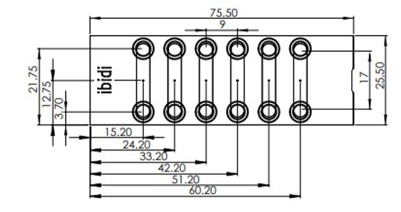
Sticky slide dimensions (mm):
*The Chitozen coverslips are made of a high-quality borosilicate glass (D 263®) with a low roughness value and a cleanroom cleaning level, specifically made for microscopy applications. They can therefore be used for super-resolution microscopy straight away without the need for any additional surface treatment procedures.
Additional resources:
> Product overview
> Technical Datasheet
> Safety Datasheet
> Certificate of Analysis - 2310
> Certificate of Analysis - 2406
> Certificate of Analysis - 2410
I used the Chitozen coverslips together with Corynebacterium glutamicum and Staphylococcus aureus. We appreciated the good adherence of the bacteria on the surface and the versatility of the tool.
Thanks to these coverslips, I've been able to observe the movements of individual cells in different motility mutants, enabling me to analyze their behavior in detail. These chitosan-coated coverslips are absolutely essential to the success of my project.
E. coli monolayers on Chitozen
© 2019 Tréguier et al. This content is distributed under the terms of the Creative Commons Attribution 4.0 International license.
Growth and division of E. coli on Chitozen, in LB medium
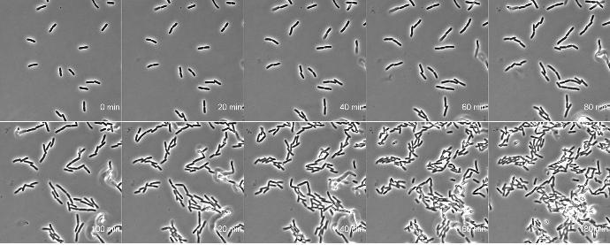
Credit: Amandine Desorme, LCB - CNRS, 2021
Monitoring amoeba motility on Chitozen
Acanthamoeba castellanii were loaded on the Chitozen slides and monitored over a 3 min time-lapse microscopy session
Credit: Flora Honoré & Tâm Mignot, LCB - CNRS, 2026
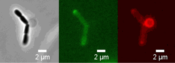
M. xanthus predates E. coli cells on Chitozen
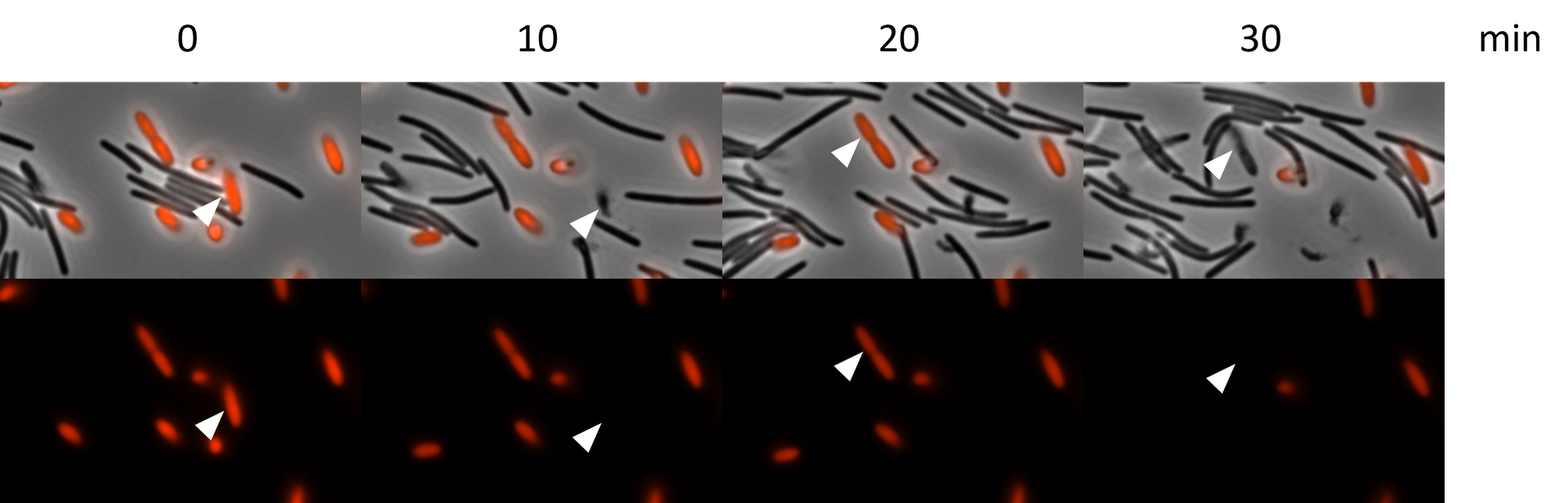
Time-lapse imaging of bacterial-bacterial interaction between M. xanthus (black) and E. Coli (red, mCherry) performed on Chitozen slide.
The white arrows show E. coli cells lysed after contact with M. xanthus cells. The experiment was conducted in CYE medium diluted 1:2 and observed on an inverted epifluorescence microscope (Nikon)
Credit: Flora Honoré and Tâm Mignot - Laboratoire de Chimie Bactérienne (LCB) - 2025
M. xanthus predates E. coli cells on Chitozen
Time-lapse imaging of bacterial-bacterial interaction between M. xanthus (black) and E. Coli (red, mCherry) performed on Chitozen slide.
The experiment was conducted in CYE medium diluted 1:2 and observed on an inverted epifluorescence microscope (Nikon)
Credit: Flora Honoré and Tâm Mignot - Laboratoire de Chimie Bactérienne (LCB) - 2025
Immunolabelling of PilA proteins in M. xanthus in predator conditions on Chitozen
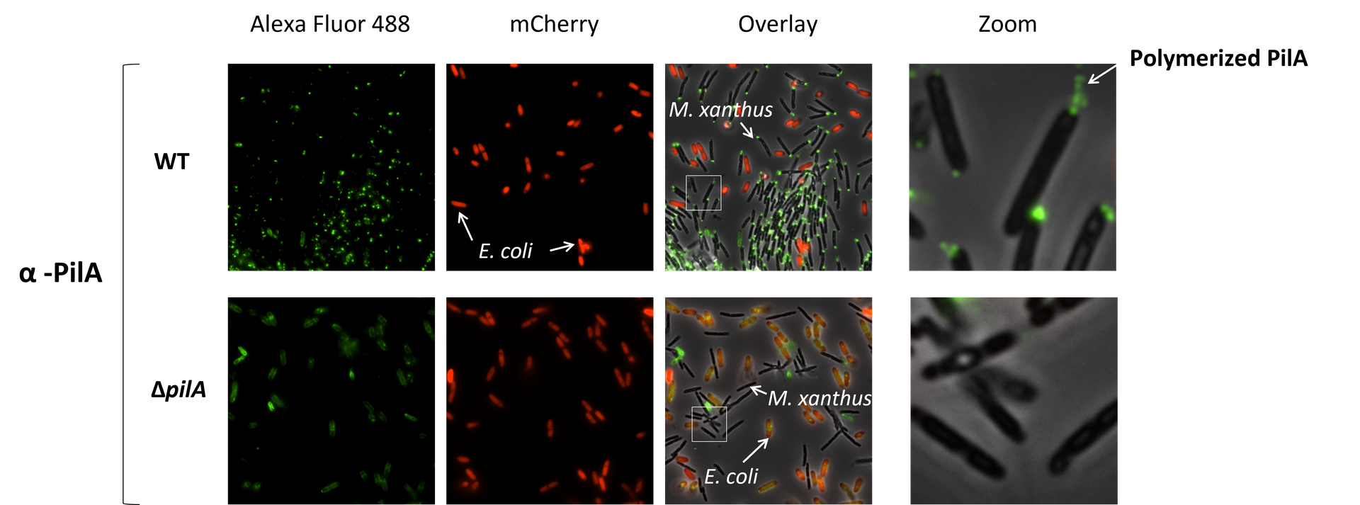
M. xanthus cells moving on chitosan are chemically fixed, then the cells are permeabilized and the PilA proteins are labeled with the PilA antibody coupled to AlexaFluor 488. PilA proteins that are polymerized extracellularly can be observed. The experiment was conducted in CYE medium diluted 1:2 and observed on an inverted epifluorescence microscope (Nikon). All fixation steps are performed directly inside the channel.
Credit: Flora Honoré and Tâm Mignot - Laboratoire de Chimie Bactérienne (LCB) - 2025
B. Exovorus digests prey content in situ on Chitozen
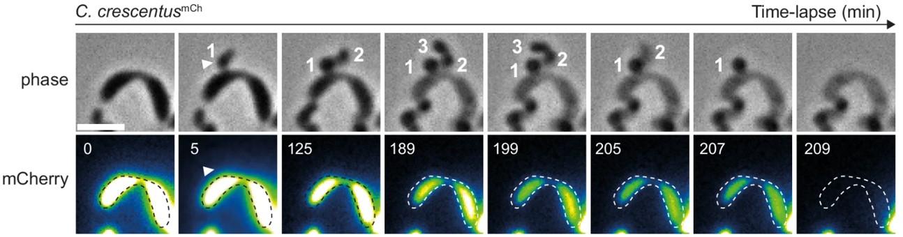
The mCherry fluorescent signal is used as a reporter of the proteinaceous cytoplasmic content. Representative time-lapse microscopy images of the mCherry-producing C. crescentus (C. crescentusmCh) predated by B. exovorus. The number of future predator daughter cells is indicated on the phase contrast images. The fluorescence signal was false colored with the GreenFireBlue colormap in Fiji to display changes in fluorescence intensity. The white arrowhead points at the B. exovorus attached to the prey surface. The predated C. crescentus cell outlines shown as dashed lines were drawn manually based on the phase contrast image at time 0. Scale bar, 2 μm
Source publication: Santin Y. et al, 2024
Visualization of Pal mCherry at septum in E. coli (W3110 Pal mCherry), by 3D SIM microscopy, in M9 medium and using Chitozen.
Credit: Amandine Desorme, LCB - CNRS, 2021
Observation of vesicles at septum during cell division of mutant E. coli (W3110 tolR - Palmcherry),
in LB 1/2 medium, using Chitozen.
Peptidoglycan is labeled with the green fluorophore BADA.
Credit: Amandine Desorme, LCB - CNRS, 2021
Time-lapse microscopy reveals kinetics of ParB condensate disassembly/reassembly
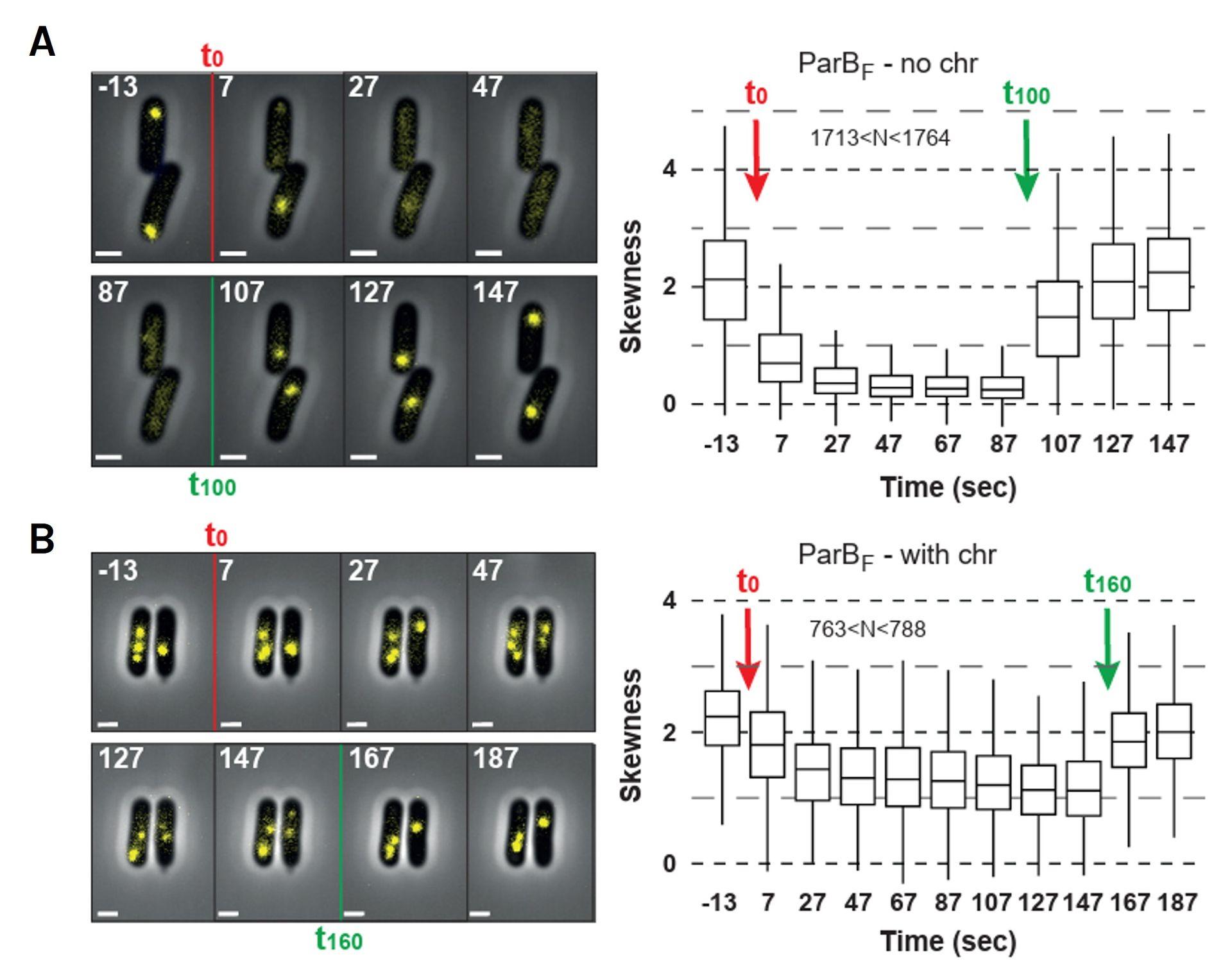
A- Rapid and reversible disassembly of ParB condensates in nucleoid-free cells. (left) Timelapse imaging of chromosome-depleted cells expressing ParBF-mVenus from plasmid F was performed using Chitozen, allowing controlled infusion of hexanediol at time 0 (T0) and wash out at 100 sec (T100). (right) Quantification of ParBF-mVenus fluorescence skewness across > 1700 cells at each indicated time point. The rapid decrease in skewness upon Hex treatment and the fast recovery upon washout indicates a nearly instantaneous disassembly and reassembly of ParB condensates. B- Rapid, reversible but partial disassembly of ParB condensates in cells with an intact chromosome. Same as in (A) but performed in cells with a nucleoid. Hex was washed out after 160 s of infusion. Fluorescence skewness was calculated from > 760 cells per time point.
Source publication: Perrine Revoil et al. Fast assembly and in vivo coalescence of ParB biocondensates involved in bacterial DNA partition. bioRxiv 2025.10.27.684735; doi: https://doi.org/10.1101/2025.10.27.684735
Live cell imaging of E. coli in response to drug addition on Chitozen slides
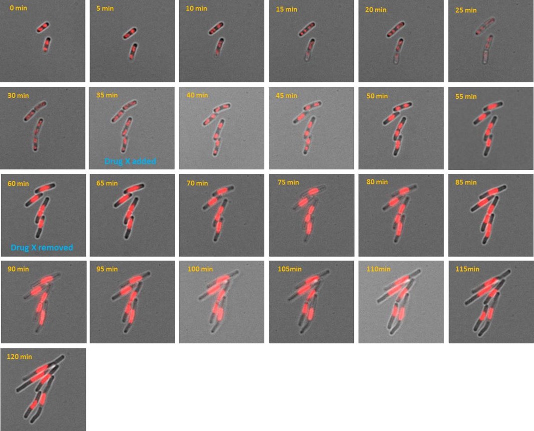
AB1157 E. coli expressing DNA marker HU-mCherry were imaged at 37oC with perfusion of ½ LB at a flow rate of 2 ml/min. Drug X was added at time point 35 minutes and removed at time point 60 minutes.
Credit: Emily Helgesen - Oslo University Hospital - 2022
Live cell imaging of E. coli expressing a protein associated with DNA replication on Chitozen slides
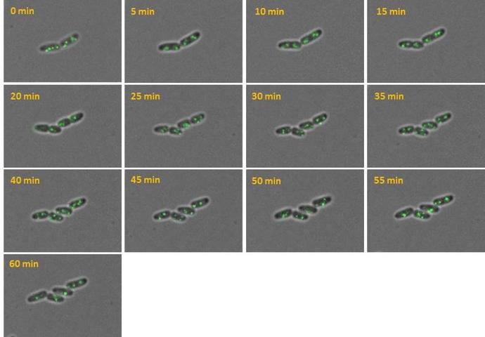
AB1157 E. coli expressing SeqA-YFP (green), a protein associated with DNA replication, were imaged at 37°C over 60 minutes without perfusion of medium
Credit: Emily Helgesen - Oslo University Hospital - 2022
Effects of cell division inhibitor cephalexin on E. coli growth cultured on Chitozen slides
E. coli BW25113 cells were imaged at 37°C with perfusion of M9 medium at a flow rate of 0.05 mL/min.
Credit: Bianca Sclavi - 2021
E. Coli spheroblasts bound to Chitozen
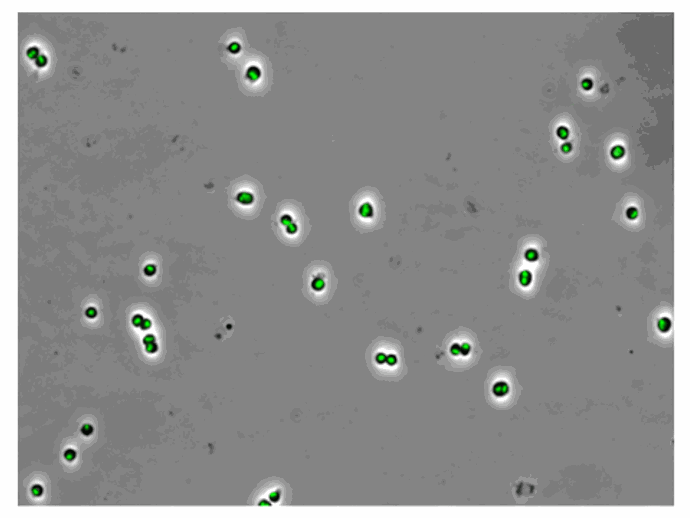
E. coli MG1655 with chromosomally encoded HU-eGFP under the native promoter were imaged with perfusion at a flow rate of 0.05 mL/min.
Credit: Itzhak Fishov – Ben-Gurion University of the Negev, 2022
DNA supercompaction in wild-type E. coli cells (KV21) after Ciprofloxacin exposure
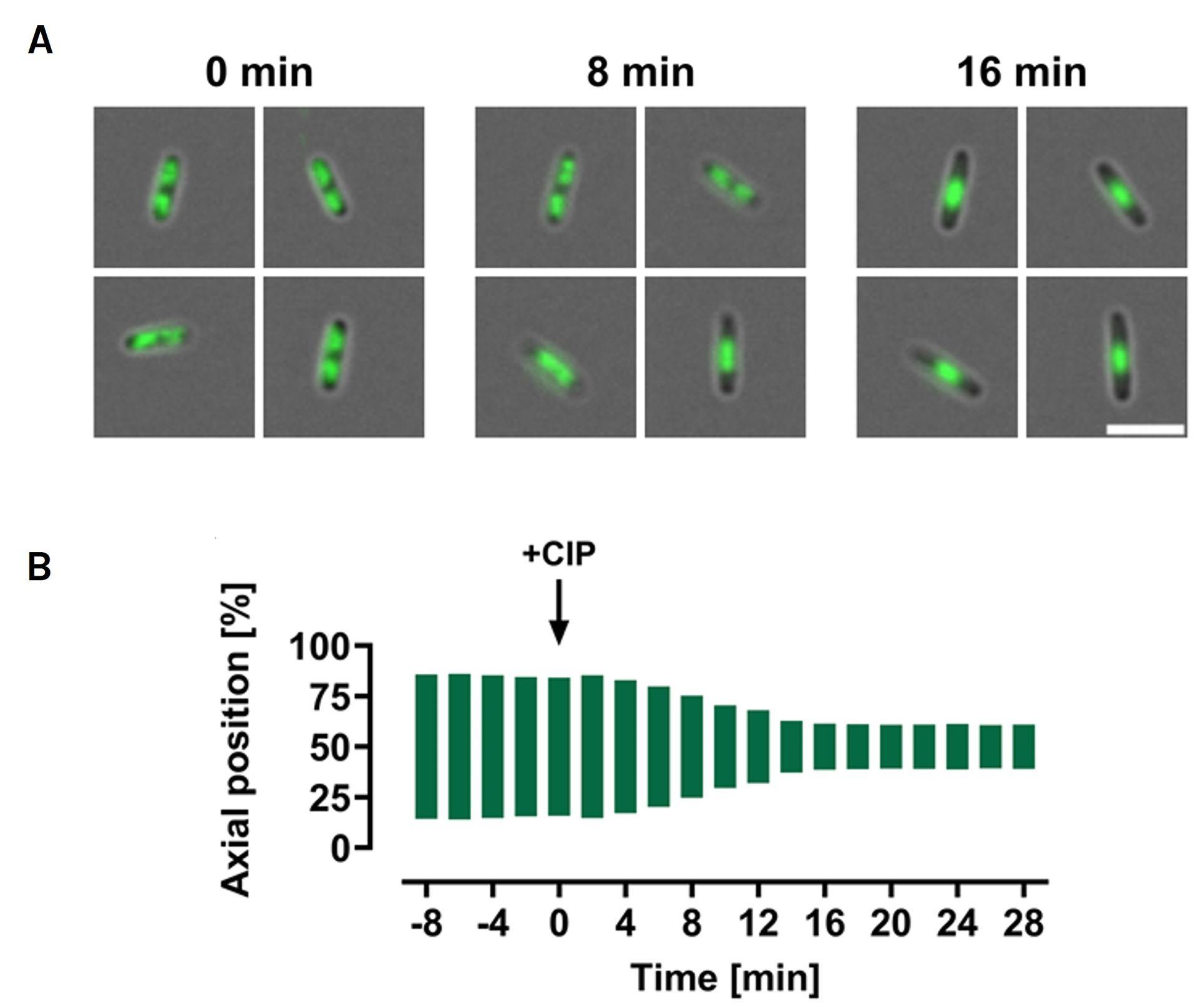
All cells were grown in LB at 37 ◦C and imaged at 2-min intervals using live-cell imaging. Cells were immobilized on Chitozen slides added with microfluidic channels and imaged using widefield microscopy. (A) Representative images of wild-type cells with HU-mCherry fluorescence (green) at 0, 8, and 16 min after CIP exposure, showing DNA distribution; scale bar: 5 μm. (B) Analysis of DNA distribution along the cells’ long axis before and after CIP, quantified by measuring the distance between the outer bounds of symmetrical fluorescence peaks at 80% of maximum a v eraged intensity for each time point. Results are averaged from 34 to 141 cells from a single representative biological replicate.
Credit: Vikedal et al, 2025
Everspark technology has been intially developed by Karine Monier, Arnaud Favier and Christophe Place and and published in Scientific Reports:
Provost, A., Rousset, C., Bourdon, L. et al. Innovative particle standards and long-lived imaging for 2D and 3D dSTORM. Sci Rep 9, 17967 (2019). https://doi.org/10.1038/s41598-019-53528-0
Publications:
Nanoscale engagement and clusterization of Programmed death ligand 1 (PD-L1) in the membrane lipid rafts of Non-Small Cell Lung Cancer cells
Martina Ruglioni, Simone Civita, Tiziano Salvadori, Sofia Cristiani, Vittoria Carnicelli, Serena Barachini, Iacopo Petrini, Irene Nepita, Marco Castello, Alberto Diaspro, Paolo Bianchini, Barbara Storti, Ranieri Bizzarri, Stefano Fogli and Romano Danesi bioRxiv 2022.08.09.503318; doi: https://doi.org/10.1101/2022.08.09.503318
HIV-1 diverts actin debranching mechanisms for particle assembly and release in CD4 T lymphocytes
Rayane Dibsy, Erwan Bremaud, Johnson Mak, Cyril Favard, Delphine Muriaux
BioRxiv, December 16, 2022. doi. 10.1101/2022.12.15.520580
Fluorescent Polymer-AS1411-Aptamer Probe for dSTORM Super-Resolution Imaging of Endogenous Nucleolin
Fabre L, Rousset C, Monier K, Da Cruz-Boisson F, Bouvet P, Charreyre MT, Delair T, Fleury E, Favier A. Biomacromolecules. 2022 May 12. doi: 10.1021/acs.biomac.1c01706. PMID: 35549176
Comparative analysis of ChAdOx1 nCoV-19 and Ad26.COV2.S SARS-CoV-2 vector vaccines.
Michalik S, Siegerist F, Palankar R, Franzke K, Schindler M, Reder A, Seifert U, Cammann C, Wesche J, Steil L, Hentschker C, Gesell-Salazar M, Reisinger E, Beer M, Endlich N, Greinacher A, Völker U. Haematologica. 2022 Apr 1;107(4):947-957. doi: 10.3324/haematol.2021.280154. PMID: 35045692
Metabolic biorthogonal labeling and dSTORM imaging of peptidoglycan synthesis in Streptococcus pneumoniae
Jennyfer Trouve, Oleksandr Glushonkov and Cecile Morlot
Star Protocols, December 13, 2021. doi: 10.1016/j.xpro.2021.101006
Insights in ChAdOx1 nCoV-19 vaccine-induced immune thrombotic thrombocytopenia.
Greinacher A, Selleng K, Palankar R, Wesche J, Handtke S, Wolff M, Aurich K, Lalk M, Methling K, Völker U, Hentschker C, Michalik S, Steil L, Reder A, Schönborn L, Beer M, Franzke K, Büttner A, Fehse B, Stavrou EX, Rangaswamy C, Mailer RK, Englert H, Frye M, Thiele T, Kochanek S, Krutzke L, Siegerist F, Endlich N, Warkentin TE, Renné T.
Blood. 2021 Dec 2;138(22):2256-2268. doi: 10.1182/blood.2021013231. PMID: 34587242
Superresolution Microscopy of Drosophila Indirect Flight Muscle Sarcomeres.
Szikora S, Novák T, Gajdos T, Erdélyi M, Mihály J. Bio Protoc. 2020 Jun 20;10(12):e3654. doi: 10.21769/BioProtoc.3654. eCollection 2020 Jun 20. PMID: 33659324