
Guy Lenaers
MitoVasc, Angers University, Angers, France
Who is
Guy Lenaers
?
Guy Lenaers is a French biomedical researcher specializing in mitochondrial genetics and neurodegenerative diseases. He serves as the Research Director at the MitoLab team within the MitoVasc Institute, affiliated with the University of Angers, CNRS (UMR 6015), and INSERM (U1083)
Dr. Lenaers is renowned for his work on inherited mitochondrial disorders, particularly those affecting the optic nerve. In 2000, his team identified the OPA1 gene, mutations of which are a primary cause of Dominant Optic Atrophy (DOA), a leading hereditary optic neuropathy. His research has significantly advanced the understanding of mitochondrial dynamics and their role in neurodegeneration.
Beyond his research, Dr. Lenaers contributes to international collaborations and serves on expert panels, such as the Optic Nerve Atrophy Variant Curation Expert Panel. His dedication to translational science aims to bridge the gap between laboratory discoveries and clinical applications, offering hope for patients with mitochondrial disorders
But beyond his fundamental research, Dr. Lenaers is also driven by a strong commitment to translational science, transforming complex discoveries into accessible, ready-to-use tools for researchers.
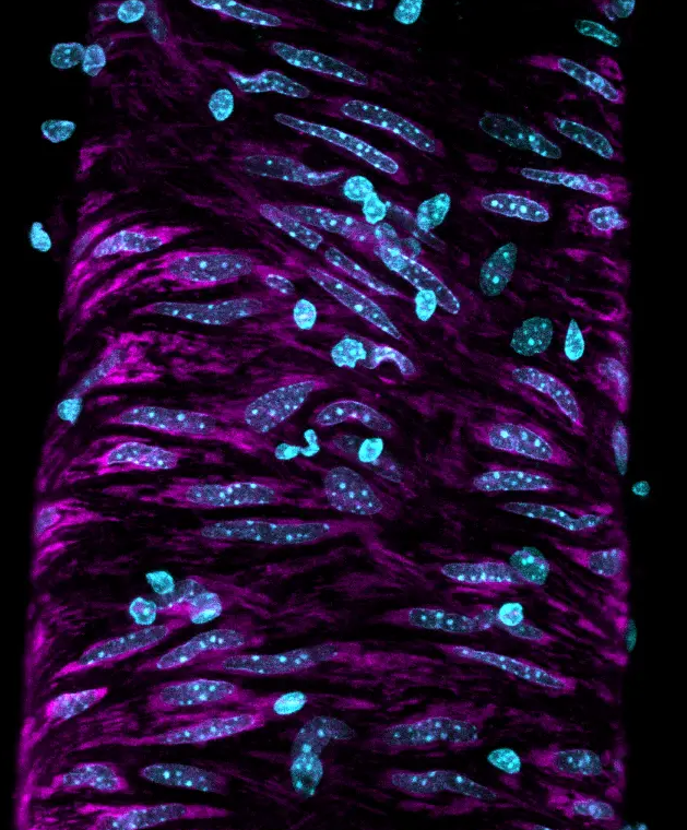
One such example is GlowMito.
GlowMito
Originally derived from the phenazine family, GlowMito was developed by Dr. Guy Lenaers and Dr. Oliver Siri
to help researchers image mitochondria without altering cell physiology , with high specificity, minimal background, and easy-to-handle protocols.
GlowMito is designed for:
- Live-cell mitochondrial tracking
- Co-labeling with other fluorophores (compatible with GFP)
- High signal retention for up to 3 days without washing
This incredible pace speaks volumes about Dr. Lenaers's vision, and his desire to transfer accessible, useful technologies to researchers everywhere.
A sibling technology: ColorFlux
When curiosity meets serendipity ..
Sometimes, breakthroughs don’t come from planned experiments, but from what happens when things don’t go as expected.
That’s exactly how ColorFlux was born.
Just four months later, ColorFlux, a bacterial efflux indicator solution, had been released by Olivier Siri’s team.
Chemists Olivier Siri and Michel Camplo were developing phenazine molecules intended to serve as antibacterial agents or adjuvants. Alongside them, Mrunal Patil, a postdoctoral pharmacist, and Jean-Michel Bolla, an expert in bacterial transport, began screening the compounds.
But the initial results? Disappointing. No strong antibacterial effect. No obvious results.
But then, something unexpected happened.
The team noticed that certain bacteria were changing color depending on their efflux activity, a subtle visual clue tied to how cells pump out foreign substances. What started as an incidental observation soon turned into a full-fledged research question:
Could these compounds be used to detect efflux behavior in bacteria?
The answer was yes, and the result is ColorFlux, a simple yet powerful colorimetric test that visually reveals bacterial efflux activity in minutes. No PCR. No fancy machines. Just color change.
 Michel Camplo Olivier Siri, Mrunal Pati et Jean-michel Bolla
Fluorescence to color change: A shared vision
ColorFlux isn’t just a colorimetric test. It’s a window into bacterial behavior, designed for any lab, no matter how high-tech. It's ideal for:
-
Quickly identifying efflux mutants and antibioresistant strains
- Evaluating potential inhibitors
- Teaching mechanisms of antibiotic resistance visually
And GlowMito continues to bring mitochondria to life under the microscope, where red fluorescence meets scientific clarity.
Two products. One molecule family. A whole lot of possibility.
ColorFlux publication
Mrunal Patil, Tatiana Munteanu, Gaël Brasseur, Carolina Ferreira, Sofia Santos Costa, Isabel Couto, Mohd Athar, Elisa Asunis, Attilio Vittorio Vargiu, Miguel Viveiros, Carole DiGiorgio, Frédéric Brunel, Jean-Manuel Raimundo, Michel Camplo, Olivier Siri, Jean-Michel Bolla —Unlocking the Gates: A Novel Diagnostic Molecule for Quantifying Efflux Levels in Gram-Positive Bacteria. doi: 10.1002/adhm.2024041451.