Biography
I am a researcher at CNRS at the University of Bordeaux (LP2N Lab) with a PhD in physics (Aix-Marseille University).I develop new optical systems for microscopy, especially applied to biology.
Research topics
I have been developing optical microscopy tools since my Master student graduation. I am fascinated by playing with the light to maximize the knowledge we can get from a sample. I am currently working in the field of super-resolution imaging assisted by robust interferometry.Fields of interest
Microscopy & imaging; light-matter interaction
Technologies
Applications
Phimask
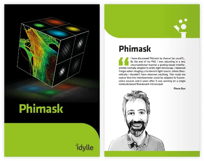
The phase-only diffraction grating designed for optimal super-resolution PSF sampling. Phimask allows 3D localization deep in tissue with a quasi-isotropic precision and limited photon loss (<20%).
Several publications
Linarès-Loyez J, Ferreira JS, Rossier O, Lounis B, Giannone G, Groc L, Cognet L and Bon P. Self-Interference (SELFI) Microscopy for Live Super-Resolution Imaging and Single Particle Tracking in 3D. Front Phys. 2019 May;7:68.
https://www.frontiersin.org/articles/10.3389/fphy.2019.00068/full
Kellermayer B, Ferreira JS, Dupuis J, Levet F, Grillo-Bosch D, Bard L, Linarès-Loyez J, Bouchet D, Choquet D, Rusakov DA, Bon P, Sibarita JB, Cognet L, Sainlos M, Carvalho AL, Groc L. Differential Nanoscale Topography and Functional Role of GluN2-NMDA Receptor Subtypes at Glutamatergic Synapses. Neuron. 2018 Oct 10;100(1):106-119.e7.
https://www.cell.com/neuron/fulltext/S0896-6273(18)30785-2?_returnURL=https%3A%2F%2Flinkinghub.elsevier.com%2Fretrieve%2Fpii%2FS0896627318307852%3Fshowall%3Dtrue
Khadir S, Bon P, Vignaud D, Galopin E, McEvoy N, McCloskey D, Monneret S, Baffou. Optical Imaging and Characterization of Graphene and Other 2D Materials Using Quantitative Phase Microscopy. ACS Photonics. 2017 Sep, 4(12):3130-3139.
https://pubs.acs.org/doi/10.1021/acsphotonics.7b00845
Aknoun S, Bon P, Savatier J, Wattellier B, Monneret S. Quantitative retardance imaging of biological samples using quadriwave lateral shearing interferometry. Opt Express. 2015 Jun 15;23(12):16383-406.
https://pubs.acs.org/doi/10.1021/acsphotonics.7b00845
Aknoun S, Savatier J, Bon P, Galland F, Abdeladim L, Wattellier B, Monneret S. Living cell dry mass measurement using quantitative phase imaging with quadriwave lateral shearing interferometry: an accuracy and sensitivity discussion. J Biomed Opt. 2015;20(12):126009.
https://www.spiedigitallibrary.org/journals/journal-of-biomedical-optics/volume-20/issue-12/126009/Living-cell-dry-mass-measurement-using-quantitative-phase-imaging-with/10.1117/1.JBO.20.12.126009.full?SSO=1
Bon P, Bourg N, Lécart S, Monneret S, Fort E, Wenger J, Lévêque-Fort S. Three-dimensional nanometre localization of nanoparticles to enhance super-resolution microscopy. Nat Commun. 2015 Jul 27;6:7764.
https://www.nature.com/articles/ncomms8764
Bourg N, Mayet C, Dupuis G, Barroca T, Bon P, Lécart S, Fort E, Lévêque-Fort S. Direct optical nanoscopy with axially localized detection. Nature Photonics. 2015 Sep ; 9(9) :587-593.
https://arxiv.org/pdf/1410.1563.pdf
Bon P, Lécart S, Fort E, Lévêque-Fort S. Fast label-free cytoskeletal network imaging in living mammalian cells. Biophys J. 2014 Apr 15;106(8):1588-95.
https://www.sciencedirect.com/science/article/pii/S000634951400229X
Baffou G, Bon P, Savatier J, Polleux J, Zhu M, Merlin M, Rigneault H, Monneret S. Thermal imaging of nanostructures by quantitative optical phase analysis. ACS Nano. 2012 Mar 27;6(3):2452-8.
https://pubs.acs.org/doi/10.1021/nn2047586

