CellAlign Myo - Myoblast alignement and differentiation
A ready-to-use substrate that guide myoblast alignment and support differentiation into long multinucleated myotubes.
A technology developed by Fouzia Boulmedais and Muhammad Haseeb Iqbal (Institut Charles Sadron, Strasbourg, France)
Designed to align. Built to differentiate.
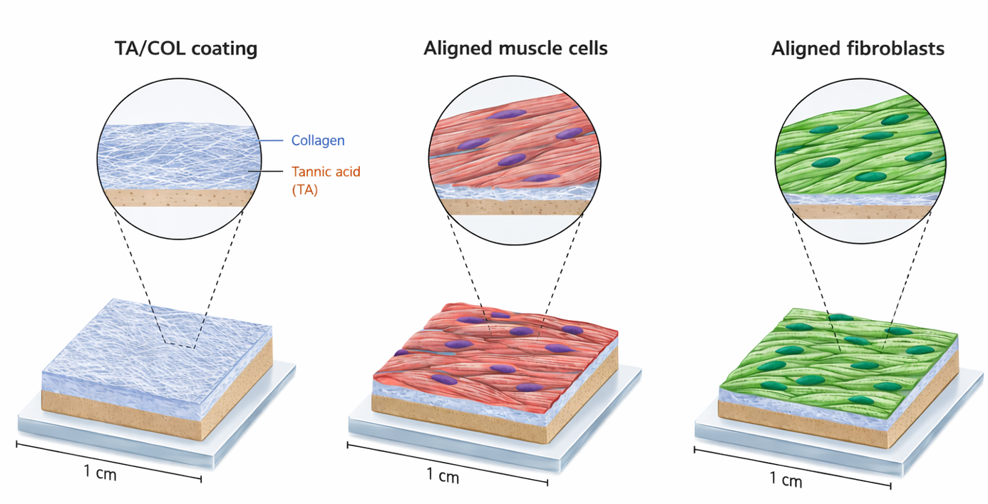
CellAlign is a pre-coated culture substrate based on oriented tannic acid / collagen films. It is designed for researchers who want a simple way to culture muscle cells on an aligned biomimetic surface. The oriented substrates guide myoblast alignment and support differentiation into long multinucleated myotubes.
CellAlign supports both structural organization and phenotypic maturation in muscle cell culture
Cellular alignment
CellAlign provides an oriented collagen-based surface that promotes the organization of myoblasts and fibroblasts and supports the formation of long, aligned myotubes.
Ready-to-use substrate
CellAlign is delivered as a finished coated substrate. Users receive a ready-to-use aligned surface, removing the need for in-house coating, patterning, or surface-preparation development.
Biocompatible
CellAlign combines collagen topographical guidance with tannic-acid-based film chemistry. Tannic acid release was linked to differentiation support, and showed local antibacterial activity.
Applications
-
Muscle cell culture
- Fibroblast cell culture
- Myoblast alignment studies
- Myotube formation assays
- Fibroblast alignment studies
- Cell morphology and focal adhesion studies
- In vitro models requiring anisotropic cell organization
* CellAlign substrates were also evaluated with fibroblasts, which adhered, spread, proliferated, and aligned on the oriented surface.
How does it work?
CellAlign works by combining directional surface guidance with bioactive TA/COL chemistry.
- The oriented collagen architecture gives cells a clear physical cue, helping them align and organize along the substrate.
- The tannic acid/collagen film chemistry supports the biological performance of the surface and contributes to the differentiation response observed in our muscle-cell model.
This combination helps create a more structured and biologically relevant culture environment than a conventional flat surface.
Validation highlights
- CellAlign Myo showed preserved nanostructure after 3 days in DMEM.
- Human myoblasts cultured on the oriented substrates formed long aligned myotubes after 12 days of differentiation.
- Tannic acid containing films showed local antibacterial activity and remained largely stable in physiological buffer.
Storage and shelf life
- Store at 4°C and protected from light
- For optimal performance, CellAlign should be used within 4 to 6 weeks of receipt
Discover the full protocol here
Download protocol
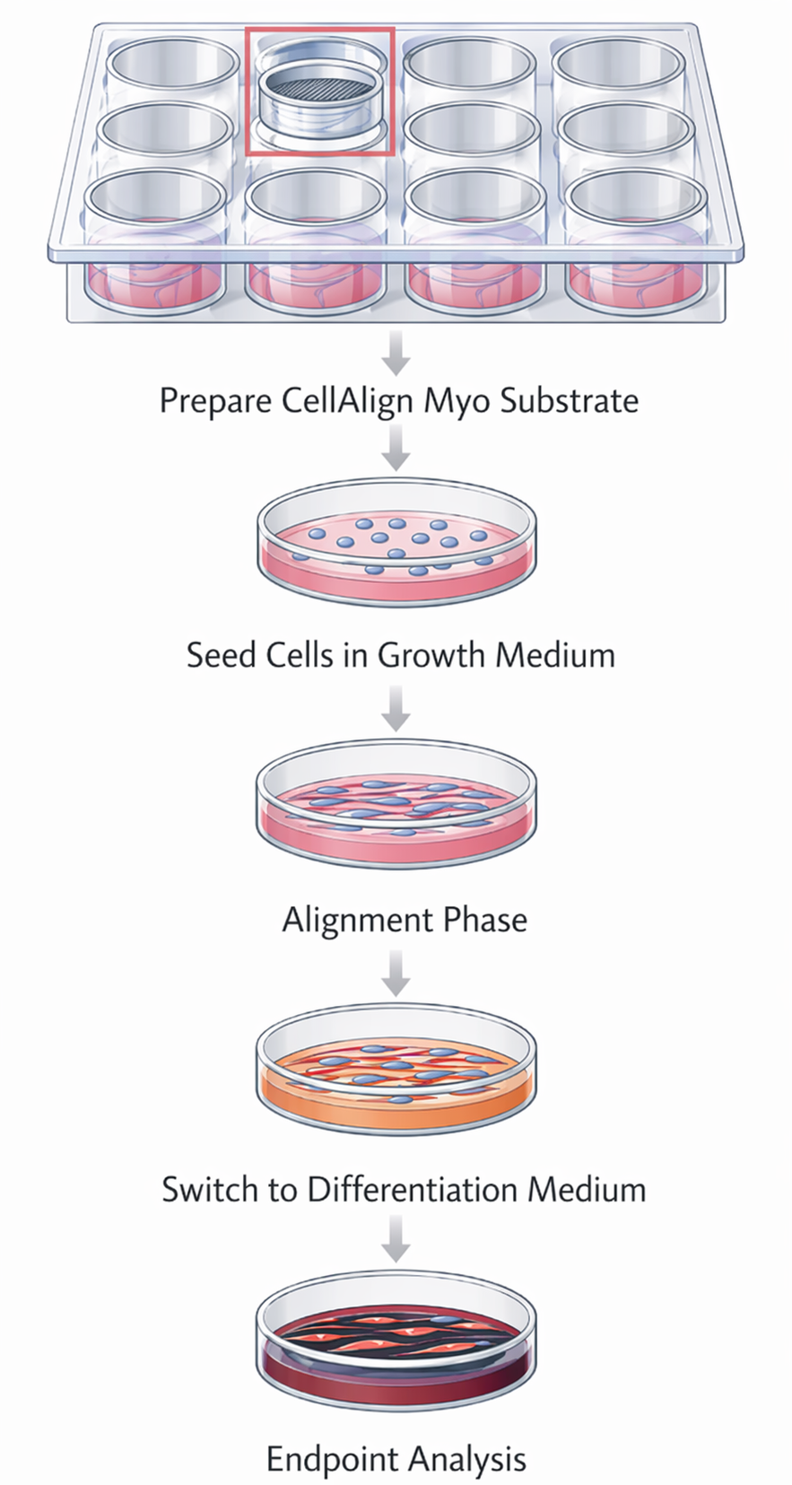
Protocol overview that shows the simplicity in adopting CellAlign substrates into your workflow:
Long, aligned human myotube formation on CellAlign Myo substrates
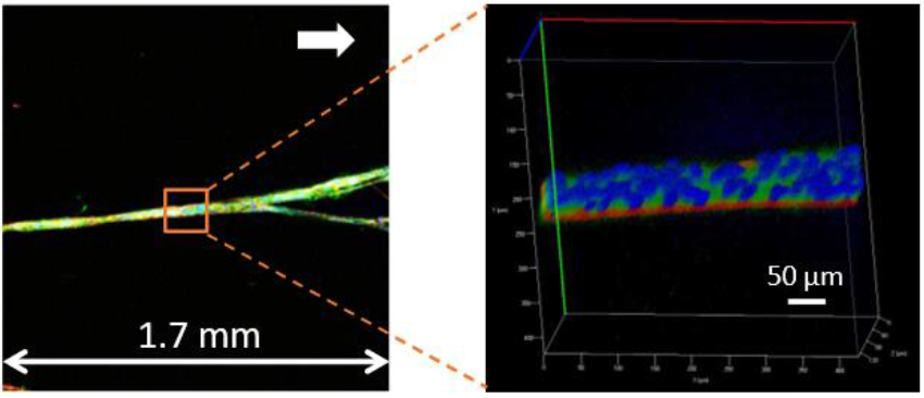
Confocal images of a long human myotube formed after 12 days of culture on CellAlign Myo substrates. The myotube extends over approximately 1.7 mm, with nuclei shown in blue, myosin heavy chain in green, and actin filaments in red. A 3D reconstructed zoom of the selected region is shown on the right.
Iqbal et al. 2022
CellAlign drives aligned myotube formation
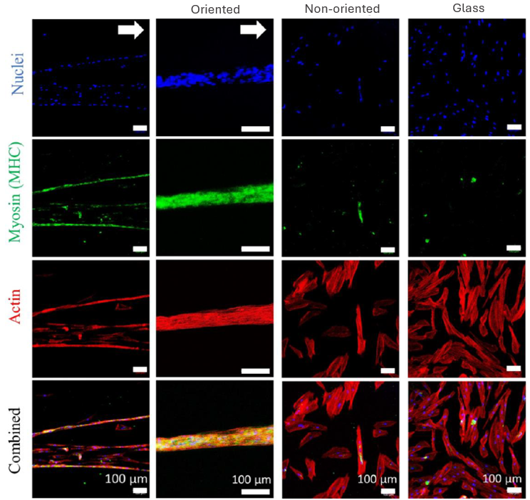
Immunostaining of human myoblast cultures highlights the biological effect of CellAlign: nuclei (blue), myosin heavy chain/MyHC (green), and actin (red) reveal the formation of long, aligned MyHC-positive myotubes on oriented substrates, compared with less organized differentiation on non-oriented substrates and glass. The white arrow marks the orientation direction of the substrate.
Iqbal et al. 2022
Myotubes elongate and align along the CellAlign orientation axis
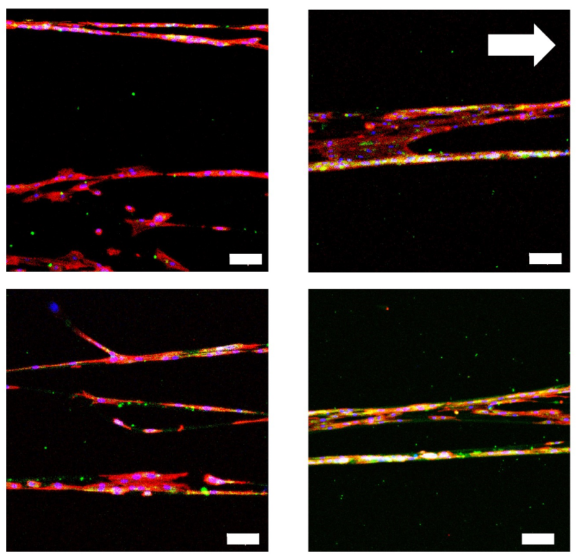
Human myoblasts form elongated, aligned MyHC-positive myotubes that follow the substrate orientation direction marked by the white arrow. Nuclei are shown in blue, myosin heavy chain (MyHC), a marker of muscle differentiation, is shown in green, and actin is shown in red.
Iqbal et al. 2022
CellAlign drives larger, longer, and more mature myotubes
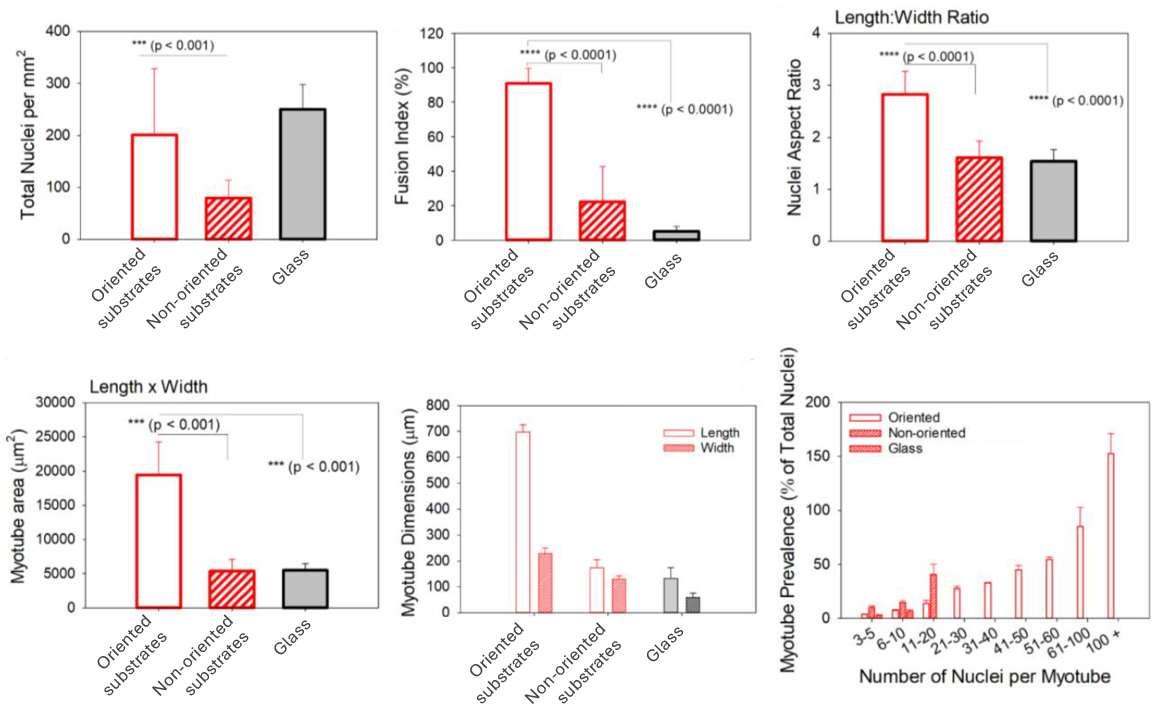
Quantitative analysis confirms the biological advantage of oriented substrates over non-oriented ones and glass controls. Compared with the other surfaces, the oriented substrates produce a much higher fusion index, more elongated nuclei, and markedly larger myotube area. They also generate longer myotubes and a much greater prevalence of highly multinucleated myotubes, indicating more advanced and organized muscle differentiation overall.
Iqbal et al. 2022
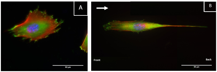
CellAlign promotes aligned and elongated cell organization
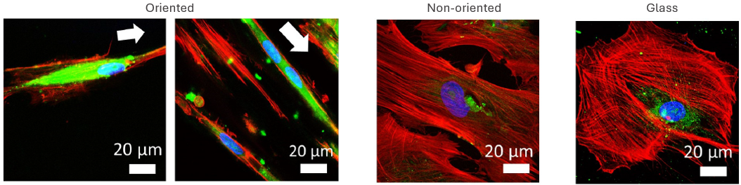
Confocal microscopy at higher magnification shows that CellAlign promotes a more elongated and directionally organized cell morphology. Nuclei are shown in blue, MyHC in green, and actin in red. The white arrows mark the substrate orientation direction, along which cells on the oriented TA/COL films become visibly more aligned than on non-oriented films or glass.
Iqbal et al. 2022
Fibroblasts cultured on CellAlign substrates showed elongated morphology and alignment along the oriented surface
Nuclei are shown in blue, actin is shown in red, and vinculin, a focal adhesion marker, is shown in green.
Credits to: Emeline Pradel, Fouzia Boulmedais and Muhammad Haseeb Iqbal
CellAlign Myo promotes aligned cell organization
During the first 48 hours, oriented TA/COL films guided myoblasts to align and cluster, which likely supported later fusion and differentiation. On non-oriented films and uncoated glass, cell movement stayed random.
Iqbal et al. 2022