- All Products
- FlashFix - Optofixation solution
FlashFix - Optofixation solution
https://www.idylle-labs.com/shop/flashfix-optofixation-solution-969 https://www.idylle-labs.com/web/image/product.template/969/image_1920?unique=4573cafSolution for cell fixation with high spatial and temporal resolution.
A technology originally developed by Stephanie Bosch (1), Maelle Carraz (1,2), Romain Duval (2)
(1) Centre de Biologie Intégrative (CBI), Unité de Biologie Moléculaire, Cellulaire et du Développement (MCD), CNRS, Toulouse, France
(2) Institut de Recherche pour le Développement (IRD), Paris , France
FlashFix, a photofixative technology.
FlashFix is a new way of fixing cell with high spatial and temporal resolution. It is based on a molecule which can trigger cell fixation when exposed to light in the proper culture conditions.
Fast fixation
Cells are snap fixed, down to 7 seconds
Precision
Resolution of fixation down to single cell
Visible
Nuclei are stained in green after photofixation
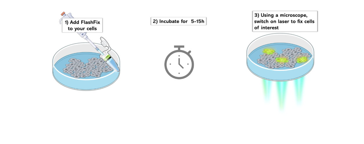
How does FlashFix work ?

Applications
- Snap fix the cells at a precise point to capture rare cellular events.
- Study the behaviour of surrounding cells when a group of cells are fixed, similarly to photo-ablation
Validated samples
FlashFix has been successfully used on a variety of cells :
- Hep3B : liver cancer cell line
- C4-2B : prostate cancer cell line
- PC3 : bone metastasis cell line of a grade IV prostatic adenocarcinoma
- T24 : urinary bladder cell line of colorectal adenocarcinoma
- WI-38 : fibroblast-like fetal lung cell.
- Cells dissociated from zebrafish embryo
Kit description:
- 1000 X stock photofixing solution
- Specific cell culture media
Find out all results linked to FlashFix in the publication section !
Long-term immobilisation of cells following optofixation with FlashFix

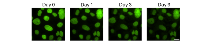
Hep3B cells were imaged right after optofixation (day 9) and subsequently followed for up to 9 days. Nuclei of fixed cells are represented in green. The absence of movement of nuclei after time shows the efficiency of fixation process. In another set of experiments, a reduction of lipid droplet movement also indicated a global absence of any intracellular movement. Scale bar = 20µm.
High control over fixation spatial resolution with FlashFix
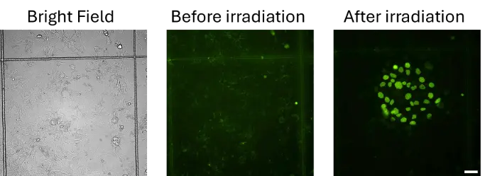
Upon illumination with 488nm light, cells were locally fixed with FlashFix. The left panel shows Hep3B cells visible in bright field. The middle and left panels show the GFP signal of cells incubated with FlashFix before (middle panel) and after (right panel) irradiation. Cells centered within the irradiation beam were strongly labeled in green, which is an indicator of their fixed status. Scale bar = 50µm.
Fixation with FlashFix is correlated with nuclear fluorescence
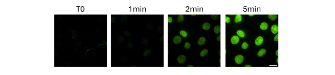
FlashFix fixative compound becomes fluorescent when it binds to DNA. Under normal culture conditions, it can partially diffuse into mitochondria and stains very faintly mitochondrial DNA. After light irradiation, the cells become permeabilized, allowing the FlashFix compound to enter the nuclei, which then emit green fluorescence. The appearance of nuclear fluorescence confirms that fixation has occurred.
Capture of precise mitotic events with FlashFix

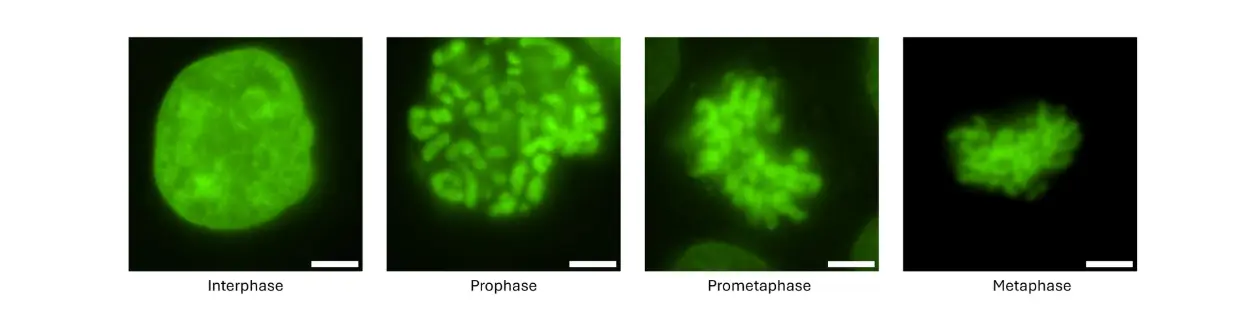
Irradiation of cells incubated with FlashFix at distinct phases of the cell cycle (Interphase, prophase, prometaphase, metaphase). Scale bar: 5 μm
Compatibility of FlashFix with immunofluorescence labeling
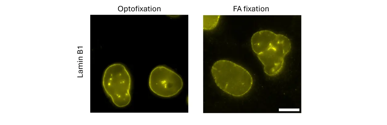
Comparison of immunofluorescence labeling following FlashFix and formaldehyde (FA) fixation. Representative image of nuclear membrane (LaminB1) staining. The marker is detectable in a similar manner in both conditions. Additional nuclear markers were successfully used, including euchromatin (H3Ac), heterochromatin (SUZ12), interspace chromatin (PML bodies), telomers (TRF1). Scale bar = 10µm
Versatility of FlashFix with multiple cell types
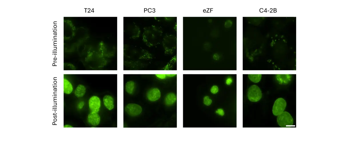
When cells are incubated with FlashFix, they will become globally fluorescent. The pattern of fluorescence can change according to the cell type and condition. Upon illumination with a 488nm laser, the fluorescence will be re-located only to the nucleus, confirming fixation. (T24 : urinary bladder of a male with colorectal adenocarcinoma; PC3 : cell line bone metastasis of a grade IV prostatic adenocarcinoma; eZF : cells dissociated from 48 hpf 5 zebrafish embryo; C4-2B : cell line prostate cancer).
Click below to download the FlashFix protocol
Upon cells’ irradiation, interaction of FlashFix components with nuclear DNA leads to singlet oxygen generation, lipid peroxidation and LPO-derived fixing aldehydes production resulting in fast (time inferior to solely formaldehyde cell-treatment) and massive intracellular crosslinks thereby, cell fixation.
Irradiation in the presence of photosensitizers usually leads to cell death through a process named photodynamic therapy. Optofixation is observed at higher irradiation intensities which kinetically overtake cell suffering and preserves morphological features of cells while quickly fixing.
Spatial and temporal resolution will depend on your microscope set up (light source power, objective numerical aperture and magnification). Classical wide field microscopes (not confocal) are suitable when equipped with a 100X magnification objective and a blue light excitation (around 488 nm). To ensure proper optofixation of cells, we recommend an irradiation power above 15W/ cm² (please refer to the protocol).
The emission spectrum of FlashFix is similar to the GFP spectrum; therefore you can combine with FlashFix other fluorescent probes or proteins before or after optofixation. Of note, these other fluorescent probes/ proteins will most likely photobleach during irradiation.
Spatial and temporal resolution will depend on your microscope set up (light source power, objective numerical aperture and magnification). Classical wide field microscopes (not confocal) are suitable when equipped with a 100X magnification objective and a blue light excitation (around 488 nm). To ensure proper optofixation of cells, we recommend an irradiation power above 15W/ cm² (please refer to the protocol).
Multiple cell islets can be optofixed within the same cell monolayer preparation and cells not targeted by light will not be fixed. At the closed proximity of optofixed cells, some phototoxicity might appear within 30 min, depending on the irradiation beam diameter.
The current version of FlashFix is not proved
yet to work on tissues or embryos. The
research team who originally developed FlashFix is currently working on an optimized
version of FlashFix which will be more suitable for tissues. If of interest for
you, please contact us for updated information.
- Original preprint behind FlashFix technology :
Light-dependent cell fixing with DNA-targeting fluorophores, BioRXiv 2026. Maëlle Carraz, Stéphanie Bosch, Thomas Mangeat, Sylvain Cantaloube, Vincent Amarh & Romain Duval (https://doi.org/10.64898/2026.03.27.714905)
Everspark technology has been intially developed by Karine Monier, Arnaud Favier and Christophe Place and and published in Scientific Reports:
Provost, A., Rousset, C., Bourdon, L. et al. Innovative particle standards and long-lived imaging for 2D and 3D dSTORM. Sci Rep 9, 17967 (2019). https://doi.org/10.1038/s41598-019-53528-0
Publications:
Nanoscale engagement and clusterization of Programmed death ligand 1 (PD-L1) in the membrane lipid rafts of Non-Small Cell Lung Cancer cells
Martina Ruglioni, Simone Civita, Tiziano Salvadori, Sofia Cristiani, Vittoria Carnicelli, Serena Barachini, Iacopo Petrini, Irene Nepita, Marco Castello, Alberto Diaspro, Paolo Bianchini, Barbara Storti, Ranieri Bizzarri, Stefano Fogli and Romano Danesi bioRxiv 2022.08.09.503318; doi: https://doi.org/10.1101/2022.08.09.503318
HIV-1 diverts actin debranching mechanisms for particle assembly and release in CD4 T lymphocytes
Rayane Dibsy, Erwan Bremaud, Johnson Mak, Cyril Favard, Delphine Muriaux
BioRxiv, December 16, 2022. doi. 10.1101/2022.12.15.520580
Fluorescent Polymer-AS1411-Aptamer Probe for dSTORM Super-Resolution Imaging of Endogenous Nucleolin
Fabre L, Rousset C, Monier K, Da Cruz-Boisson F, Bouvet P, Charreyre MT, Delair T, Fleury E, Favier A. Biomacromolecules. 2022 May 12. doi: 10.1021/acs.biomac.1c01706. PMID: 35549176
Comparative analysis of ChAdOx1 nCoV-19 and Ad26.COV2.S SARS-CoV-2 vector vaccines.
Michalik S, Siegerist F, Palankar R, Franzke K, Schindler M, Reder A, Seifert U, Cammann C, Wesche J, Steil L, Hentschker C, Gesell-Salazar M, Reisinger E, Beer M, Endlich N, Greinacher A, Völker U. Haematologica. 2022 Apr 1;107(4):947-957. doi: 10.3324/haematol.2021.280154. PMID: 35045692
Metabolic biorthogonal labeling and dSTORM imaging of peptidoglycan synthesis in Streptococcus pneumoniae
Jennyfer Trouve, Oleksandr Glushonkov and Cecile Morlot
Star Protocols, December 13, 2021. doi: 10.1016/j.xpro.2021.101006
Insights in ChAdOx1 nCoV-19 vaccine-induced immune thrombotic thrombocytopenia.
Greinacher A, Selleng K, Palankar R, Wesche J, Handtke S, Wolff M, Aurich K, Lalk M, Methling K, Völker U, Hentschker C, Michalik S, Steil L, Reder A, Schönborn L, Beer M, Franzke K, Büttner A, Fehse B, Stavrou EX, Rangaswamy C, Mailer RK, Englert H, Frye M, Thiele T, Kochanek S, Krutzke L, Siegerist F, Endlich N, Warkentin TE, Renné T.
Blood. 2021 Dec 2;138(22):2256-2268. doi: 10.1182/blood.2021013231. PMID: 34587242
Superresolution Microscopy of Drosophila Indirect Flight Muscle Sarcomeres.
Szikora S, Novák T, Gajdos T, Erdélyi M, Mihály J. Bio Protoc. 2020 Jun 20;10(12):e3654. doi: 10.21769/BioProtoc.3654. eCollection 2020 Jun 20. PMID: 33659324