The Luminicell Trackers™ are a novel family of ultra-bright, long-lived fluorescent cytosolic markers with low cytotoxicity for in vitro & in vivo long-term live cell tracking. Once internalized, the labelled dyes can be tracked up to 10 generations in vitro and up to 6 weeks in vivo.
Extending cell tracking time: thanks to excellent cell retention properties, cells remain fluorescently tagged for up to 10 generations in vitro and 6 weeks after in vivo transplantation with no cross-talk, unlocking new potential for long-term monitoring of cell fate & distribution.
Low toxicity: >95% cell viability after 48h and no disruption of normal cell functions.
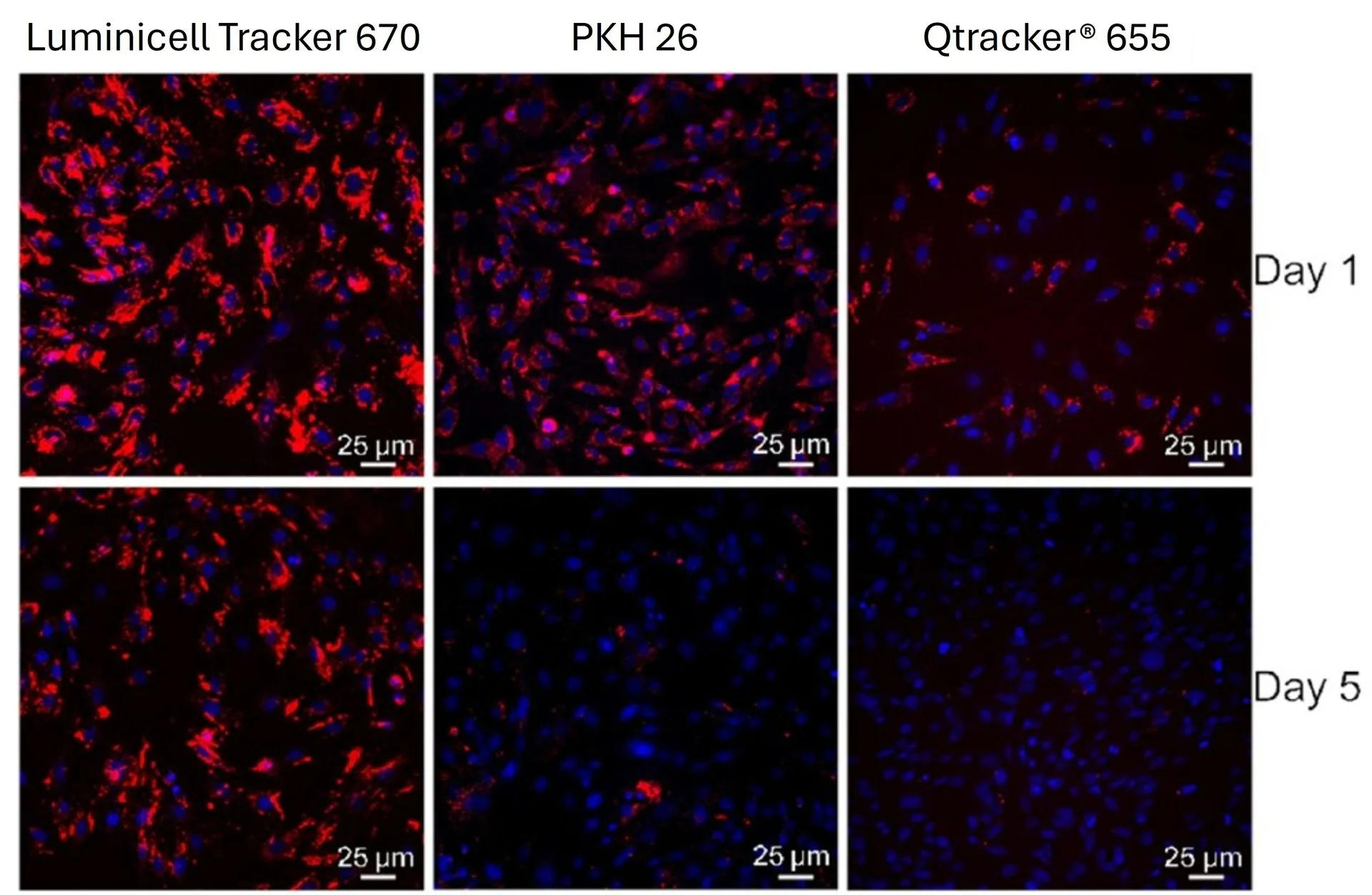
High brightness & photostability: >10X fluorescent intensity compared to quantum dots, low background auto-fluorescence and >90% fluorescence signal retention after 30 min of continuous laser excitation.
Fast & easy: simply add
Luminicell Tracker™
to your cells and get a >99% labeling rate in less than an hour. Thanks to their simple protocol, LuminiCell trackers do not require any transfection step and can be easily integrated within your existing workflow.
Versatile: with their excellent biocompatibility, deep penetration depth and high compatibility with multiphoton microscopy,
Luminicell Tracker™
will allow you to perform long-term cell tracking in a variety of samples ranging from 2D & 3D cell cultures, live tissues to small animal models.
Luminicell Tracker™ - Cell Labelling Kits are available as blue, green, red, NIR-I & NIR-II fluorescent compounds to match biological optical window.
How does Luminicell Trackers work?
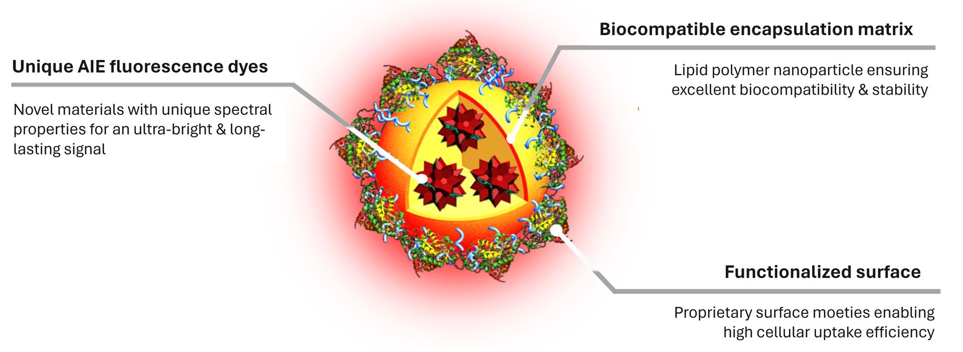
Luminicell Trackers™ are organic fluorescent nanoparticles. They are composed of unique Aggregation Induced Emission (AIE) dyes, at the heart of
Luminicell Tracker™
technology, encapsulated into lipid nanoparticles with proprietary surface. This novel class of materials emit fluorescence in an opposite manner compared to conventional fluorophores (quantum dots, GFP, etc). These unique spectral properties protect them from aggregation-caused quenching, resulting in high signal-to-noise ratio and strong photobleaching resistance, making them optimal candidates for long-term live cell tracking experiments.
Luminicell Tracker™ shows excellent biocompatibility and long-term cell retention properties for in vitro & in vivo applications. Once internalized, Luminicell Trackers™ provide an even labelling of the cytoplasmic matrix retained for up to 10 cell generations.
Find out more about the patented Luminicell Tracker™ proprietary technology platform and its well-recognized track records in our Publications section.
Applications of Luminicell Trackers
The low toxicity and long-lasting nature of Luminicell Tracker™ signal allow a wide variety of cell tracking applications both in vitro and in small animal models (i.e. cell distribution, mobility, cell-cell interactions, etc) to suit your studies from fundamental research to translational & pre-clinical studies.
Sample types:
Luminicell Trackers™
show efficient labelling rate (>99%) in a wide range of adherent & suspension cell lines and primary cells, live & fixed. Their deep penetration depth makes
Luminicell Trackers™
suitable for labelling 3D cell culture models and tissues.
Check out our FAQ section for a full list of validated samples extracted from selected publications.
Experimental outputs:
Once labelled, cells can be imaged in vitro and will remain fluorescently tagged
for up to 10 generations.
Luminicell Trackers™
of different colors can be multiplexed for use in co-cultures or 3D models with no cross-talk.
Cells labelled with
Luminicell Tracker™
can be injected into small animal models for long-term in vivo live cell tracking (up to 42 days), or subsequent ex vivo imaging of extracted organs or tissue sections. So far, they have been successfully used for cell tracking in mice and 3 dpf zebrafish.
Compatible imaging modes: Luminicell Tracker™ labeled cells can be monitored using a variety of fluorescence techniques such as epifluorescence & confocal microscopes, in vivo imaging systems (i.e. IVIS Spectrum) and flow cytometry.
Luminicell Tracker™ is compatible with multiphoton excitation microscopy. Five different versions of
Luminicell Tracker™
with blue, cyan, green, red, NIR-I & NIR-II emission colours are available for purchase through our current catalog.
Check out our Technical Specifications section for full details about optical properties and compatible instrument parameters.
Case studies: Among other applications,
Luminicell Trackers™
have been successfully used in regenerative therapy and immunotherapy development to monitor retention and biodistribution of therapeutic/engrafted cells at the single-cell resolution, tissue and whole organism scale.
Discover more about these case studies and many more in our Results and Publications sections.
Additional resources
Discover more about Luminicell trackers and their use for long-term live cell imaging with this on-demand seminar:
The
Luminicell Tracker™
probes are provided in various emission colors. Choose among our available catalog to fit your desired applications:
Luminicell Tracker™ 1010 - Cell Labelling Kit (NIR-II)
Optical properties:
Absorption max = 725 nm
Emission max = 1010 nm
Compatible instrument parameters:
Laser excitation λ (nm): 355/405/633/755*
Filter sets (nm): > 1000
Laser excitation λ (nm) - Two-photon: 1400
*best excitation wavelength for fluorescent signal
-
Concentration: 200 nM in ultrapure water
Pack size:
- Trial vial (up to 20 assays): 1x 100 µL
- Standard vial (up to 50 assays): 1x 250 µL
Luminicell Tracker™ 810 - Cell Labelling Kit (NIR-I)
Optical properties:
Absorption max = 635 nm
Emission max = 810 nm
Compatible instrument parameters:
Laser excitation λ (nm): 355/543/633*/755
Filter sets (nm): 700 - 1000
Laser excitation λ (nm) - Two-photon: 1200
*best excitation wavelength for fluorescent signal
-
Concentration: 200 nM in ultrapure water
Pack size:
- Trial vial (up to 20 assays): 1x 100 µL
- Standard vial (up to 50 assays): 1x 250 µL
Luminicell Tracker™ 670 - Cell Labelling Kit (Red)
Optical properties:
Absorption max = 506 nm
Emission max = 670 nm
Compatible instrument parameters:
Laser excitation λ (nm): 355/458/488*/543
Filter sets (nm): 670 - 800
Laser excitation λ (nm) - Two-photon: 820*/840/920
*best excitation wavelength for fluorescent signal
-
Concentration: 200 nM in ultrapure water
Pack size:
- Trial vial (up to 20 assays): 1x 100 µL
- Standard vial (up to 50 assays): 1x 250 µL
Luminicell Tracker™ 540 - Cell Labelling Kit (Green)
Optical properties:
Absorption max = 423 nm
Emission max = 540 nm
Compatible instrument parameters:
Laser excitation λ (nm): 355/405*/458/488
Filter sets (nm): 480 - 560
Laser excitation λ (nm) - Two-photon: 800
*best excitation wavelength for fluorescent signal
- Concentration: 200 nM in ultrapure water
Pack size:
- Trial vial (up to 20 assays): 1x 100 µL
- Standard vial (up to 50 assays): 1x 250 µL
Luminicell Tracker™ 470 - Cell Labelling Kit (Blue)
Optical properties:
Absorption max = 355 nm
Emission max = 470 nm
Compatible instrument parameters:
Laser excitation λ (nm): 355*/390
Filter sets (nm): 400 - 500
*best excitation wavelength for fluorescent signal
- Concentration: 200 nM in ultrapure water
Pack size:
- Trial vial (up to 20 assays): 1x 100 µL
- Standard vial (up to 50 assays): 1x 250 µL
Luminicell Tracker™ 506 - Cell Labelling Kit (Cyan)
Optical properties:
Absorption max = 355 nm
Emission max = 506 nm
Compatible instrument parameters:
Laser excitation λ (nm): 355*/405
Filter sets (nm): 420 - 520
*best excitation wavelength for fluorescent signal
- Concentration: 200 nM in ultrapure water
Pack size:
- Trial vial (up to 20 assays): 1x 100 µL
- Standard vial (up to 50 assays): 1x 250 µL
Storage: Luminicell Tracker™ probes can be stored at 2-8°C for up to 18 months. Do not freeze.
Each 250 µL vial provided in the Luminicell Tracker™ - Cell Labelling Kit allows for 25 mL total imaging medium when used at the recommended 2 nM concentration.
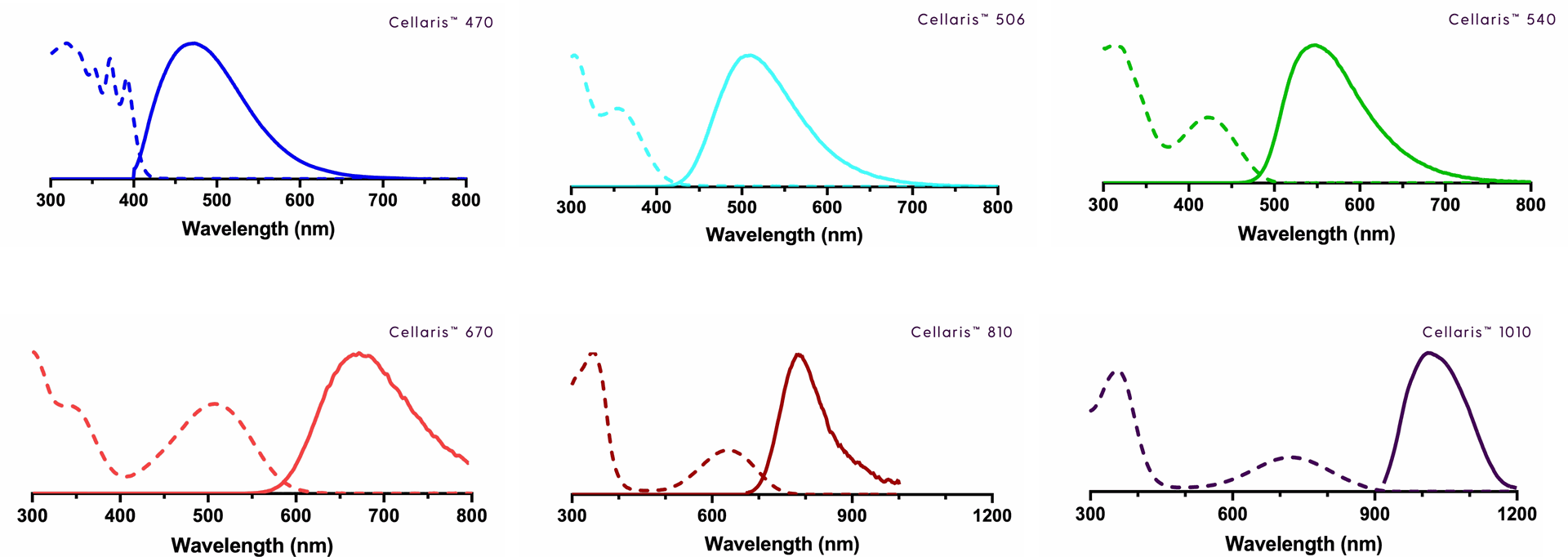
UV-vis absorption (dashed) and emission (solid) spectra of Luminicell Trackers in water
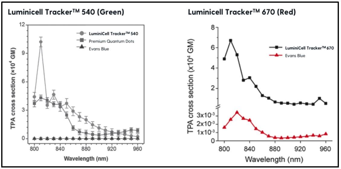
Two-photon absorption spectra of Luminicell Tracker 540 (green) and Luminicell Tracker 670 (red)
Enhanced capabilities of Luminicell Trackers for long-term tracking of adipose-derived stem cells
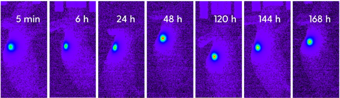
Tumour monitoring using NIR-II with Luminicell Trackers 1010
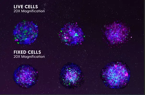
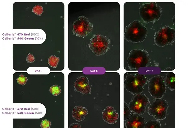
Assessing cell distribution during 3D spheroid growth using Luminicell Trackers
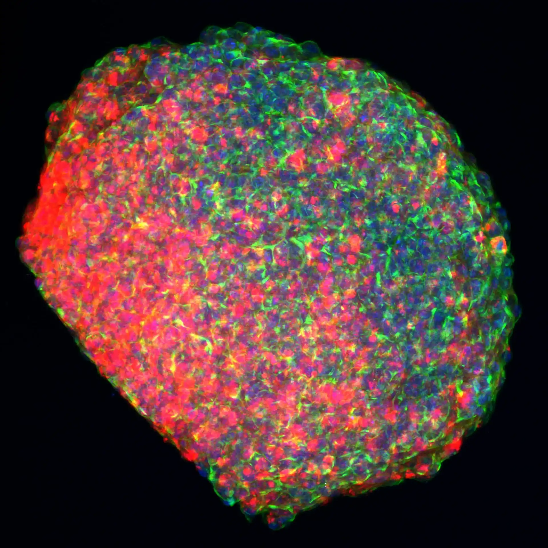
Credits: Yasemin Geiger and Dr. Stefanie Sudhop, CANTER Lab (University of Applied Sciences, Munich).
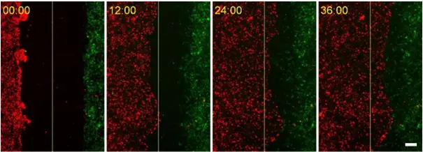
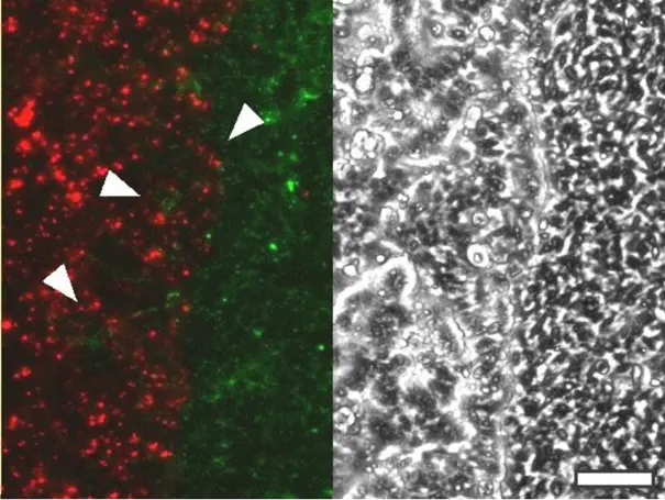
Luminicell Trackers allow clear distinction between different cell types in co-culture invasion assays
Luminicell Trackers exhibits no cross-talk effects in multi-colored experiments
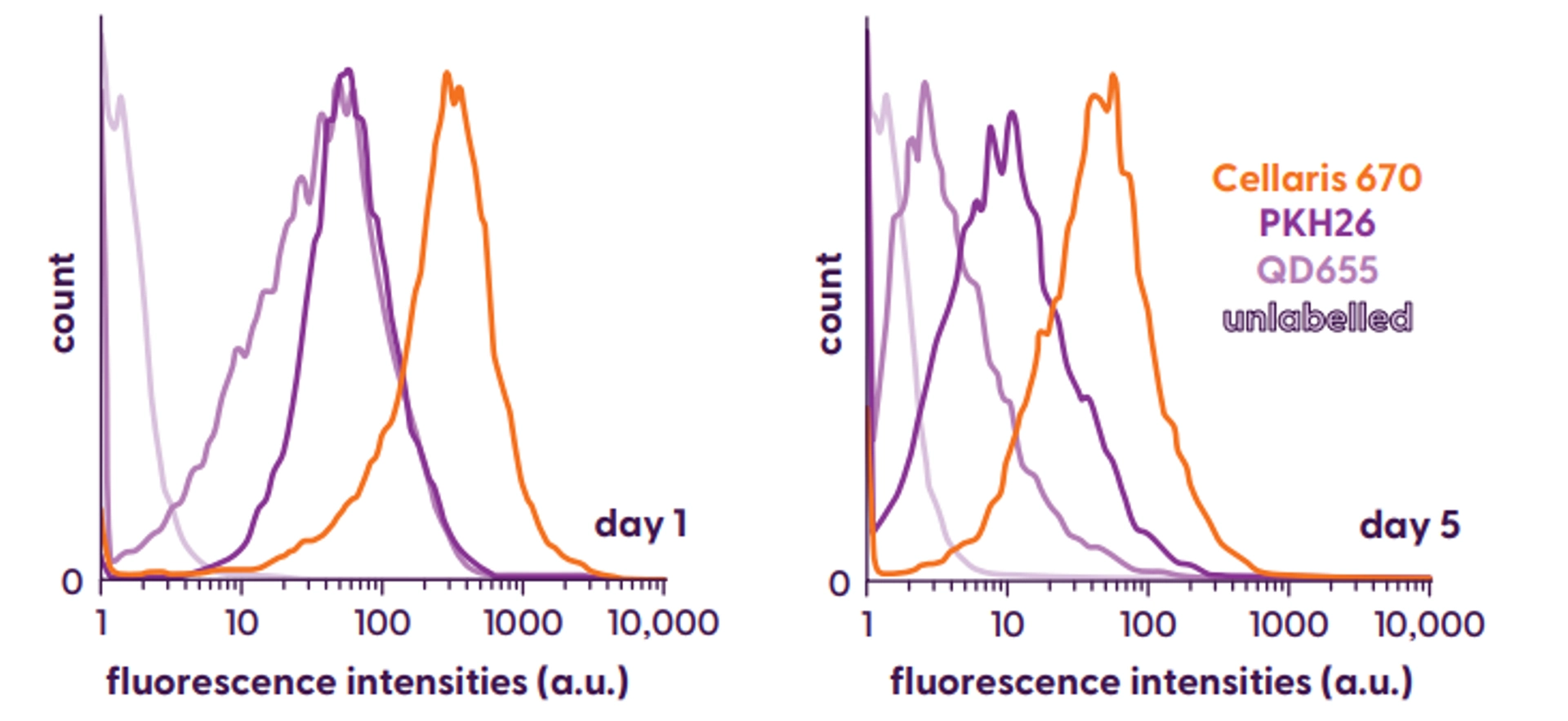
Luminicell Trackers exhibit higher fluorescence intensity and longer labelling than other tracking dyes for flow cytometry analysis
Long-term in vivo tracking of transplanted stem cells using Luminicell Trackers
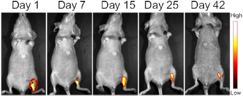
In vivo tracking of transplanted stem cells at single-cell resolution
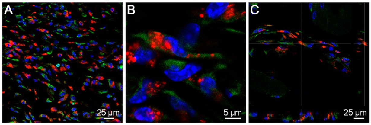
Source publication: Dan Ding et al. (2014). Precise and Long-Term Tracking of Adipose-Derived Stem Cells and Their Regenerative Capacity via Superb Bright and Stable Organic Nanodots. ACS Nano 8 (12), 12620-12631. https://doi.org/10.1021/nn505554y
Multicellular 3D Blood-Brain Barrier (BBB) model
Source: Multicellular 3D Blood-Brain Barrier (BBB) model (see Publication section)
In vitro 3D tissue culture model of fibrotic liver
Organoids incubated with ClinoStar®.
Source: In vitro 3D tissue culture model of fibrotic liver (see Publication section)
Other products you may like
Agarsqueezer
Use the AgarSqueezer confiner to facilitate your single-cell tracking experiments in 3D cell structures.
Stencell
Multiply your experimental conditions to save Luminicell tracker volumes, or use it for live cell tracking during migration.
SpheroTribe
Boost your 3D structure uniformity to simplify your cell tracking assay analysis and get the most reliable data.
Stampwell - zebrafish
Use Stampwell to facilitate imaging of labelled cells in multiple zebrafishes at the same time by immobilizing and aligning them perfectly.
Stampwell - 3D cell structures
Use Stampwell to facilitate imaging of labelled cells in multiple 3D structures at the same time by immobilizing and aligning them perfectly.
GlowMito
Combine Luminicell trackers with GlowMito, also compatible with multiphoton microscopy, and visualize mitochondria during your cell tracking experiments in living cells and thick tissues.