The Luminicell Trackers™ are a novel family of ultra-bright, long-lived fluorescent nanoparticles with low toxicity for real-time imaging of deep vasculature in live animals. Once injected in vivo, these nanoparticles flow smoothly inside blood vessels and only leak out in response to blood vessel permeability alterations upon inflammation or infection.
Compatible with multiphoton microscopy: thanks to their high two-photon absorption cross-section,
Luminicell Trackers™
are a solution of choice for deep-tissue & multiphoton intravital imaging.
Non-toxic: encapsulated into a fully biocompatible matrix for safe in vivo delivery with low toxicity and minimal biological interference [1].
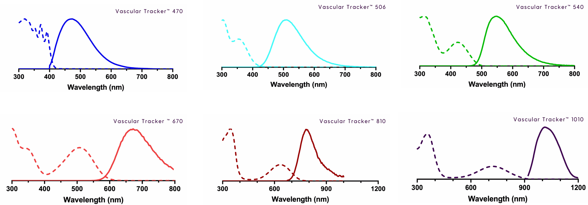
Luminicell Trackers™ - Vascular Labelling kits are available as blue, green, red, NIR-I & NIR-II fluorescent compounds to match biological optical window.
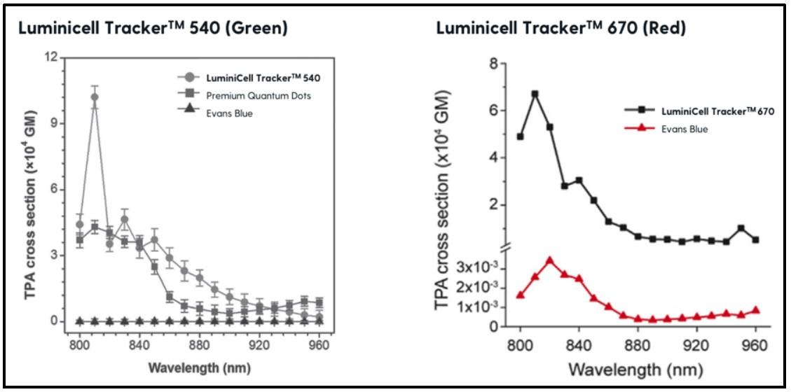
Enhanced signal: ultra-bright intrinsic fluorescence (20X higher intensity compared to Evans blue [2]) and strong photobleaching resistance after 30 min of continuous laser excitation, for increased spatial resolution and facilitated data analysis.
Excellent retention: Luminicell trackers are well-retained within intact vessels and only leak out in response to changes in permeability. Be assured your qualitative and quantitative analyses of changes in vascular functions are specific and reliable!
How does Luminicell Trackers work?
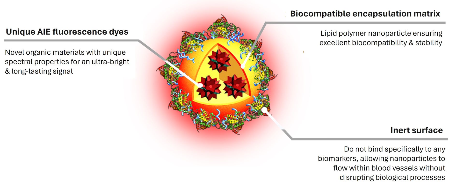
Luminicell Trackers™ are organic fluorescent nanoparticles. The are composed of unique Aggregation Induced Emission (AIE) dyes, at the heart of Luminicell Tracker™ technology, encapsulated into lipid nanoparticles with proprietary surface. This novel class of materials emit fluorescence in an opposite manner compared to conventional fluorophores (quantum dots, GFP, etc). These unique spectral properties protect them from aggregation-caused quenching, resulting in high signal-to-noise ratio and strong photobleaching resistance, making them optimal candidates for in vivo experiments.
Luminicell
Tracker™ – Vascular Labelling Kit is designed with inert surface groups, which does not bind specifically to any biomarkers, allowing them to flow smoothly inside blood vessels. Under inflammation or infection, they leak out in response to blood vessel permeability changes and form localized punctate aggregates, accumulating at surrounding tissues.
Find out more about the patented Luminicell Tracker™ proprietary technology platform and its well-recognized track records in our Publications section.
Applications of Luminicell Trackers for Vascular Labelling
The
Luminicell Tracker™
- Vascular Labelling Kits can be used to fluorescently tag vasculature in live animals for your study in vascular-related diseases and cancers.
Sample types: Luminicell Trackers can be injected in small animals (intraveneous or retro-orbital injection) or inside in vitro barrier models (i.e. 3D microfluidic devices).
Experimental outputs:
Once the Luminicell Trackers are injected in vivo, fluorescent blood vessels can be visualized in vivo using intravital live imaging systems, or ex vivo after harvesting particular organs or tissues. In both cases, the trackers can be used to:
- Visualize vasculature morphology, dynamics & perform 3D reconstructions of vascular systems
- Detect and quantify vascular leakage and changes in vascular functions
Compatible imaging modes: compatible with one & two-photon fluorescence microscopy. Six different versions of Luminicell Tracker™ with blue, cyan, green, red, NIR-I & NIR-II emission colours are available for purchase through our current catalog.
Case studies:
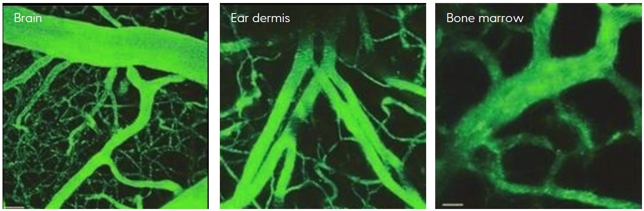
Luminicell Trackers™have been successfully used to visualize vasculature in mice brain, bone marrow, ear skin and tumors, and zebrafish brain.
Among other applications, they proved useful to detect haemorrhage at blood-brain-barrier malfunction sites in cerebral malaria or assess changes in tumor vessel structures by deep tumor imaging. They have also been used for assessing vessel permeability in a 3D microfluidic model of human micro-vessels.
Discover more about these case studies and many more in our Results and Publications sections.
Additional resources
- Vascular Tracker - Technical Brochure
- Safety Data Sheet - Luminicell Tracker™ 540 (Green)
- Safety Data Sheet - Luminicell Tracker™ 670 (Red)
References:
[1]
Feng, G. et al. (2018). Dual Modal Ultra-Bright Nanodots with Aggregation-Induced Emission and Gadolinium-Chelation for Vascular Integrity and Leakage Detection. Biomaterials. Jan;152:77-85. doi: 10.1016/j.biomaterials.2017.10.031. Epub 2017 Oct 20. PMID: 29111495.
[2] Ding, D. et al. (2013). Ultrabright Organic Dots with Aggregation-Induced Emission Characteristics for Real-Time Two-Photon Intravital Vasculature Imaging. Advanced Materials, 25(42), 6083-6088. https://doi.org/10.1002/adma.201301938
The Luminicell Tracker probes are provided in various emission colors. Choose among our available catalog to fit your desired applications:
Luminicell Tracker™ 1010 - Vascular Labelling Kit (NIR-II)
Optical properties:
Absorption max = 725 nm
Emission max = 1010 nm
-
Compatible instrument parameters:
Laser excitation λ (nm): 355/405/633/755*
Filter sets (nm): > 1000
Laser excitation λ (nm) - Two-photon: 1400
*best excitation wavelength for fluorescent signal -
Concentration: 200 nM in ultrapure water
Pack size:
- Trial vial: 1x 100 µL
- Standard vial: 1x 250 µL
Luminicell Tracker™ 810 - Vascular Labelling Kit (NIR-I)
Optical properties:
Absorption max = 635 nm
Emission max = 810 nm
-
Compatible instrument parameters:
Laser excitation λ (nm): 355/543/633*/755
Filter sets (nm): 700 - 1000
Laser excitation λ (nm) - Two-photon: 1200
*best excitation wavelength for fluorescent signal -
Concentration: 200 nM in ultrapure water
Pack size:
- Trial vial: 1x 100 µL
- Standard vial: 1x 250 µL
Luminicell Tracker™ 670 - Vascular Labelling Kit (Red)
Optical properties:
Absorption max = 506 nm
Emission max = 670 nm
-
Compatible instrument parameters:
Laser excitation λ (nm): 355/458/488*/543
Filter sets (nm): 670-800
Laser excitation λ (nm) - Two-photon: 820*/840/920
*best excitation wavelength for fluorescent signal -
Concentration: 200 nM in ultrapure water
Pack size:
- Trial vial: 1x 100 µL
- Standard vial: 1x 250 µL
Luminicell Tracker™ 540 - Vascular Labelling Kit (Green)
Optical properties:
Absorption max = 423 nm
Emission max = 540 nm
- Compatible instrument parameters:
Laser excitation λ (nm): 355/405*/458/488
Filter set (nm): 480 - 560
Laser excitation λ (nm) - Two-photon: 800
*best excitation wavelength for fluorescent signal - Concentration: 200 nM in ultrapure water
Pack size:
- Trial vial: 1x 100 µL
- Standard vial: 1x 250 µL
Luminicell Tracker™ 506 - Vascular Labelling Kit (Cyan)
Optical properties:
Absorption max = 355 nm
Emission max = 506 nm
- Compatible instrument parameters:
Laser excitation λ (nm): 355*/405
Filter sets (nm): 420 - 520
*best excitation wavelength for fluorescent signal - Concentration: 200 nM in ultrapure water
Pack size:
- Trial vial: 1x 100 µL
- Standard vial: 1x 250 µL
Luminicell Tracker™ 470 - Vascular Labelling Kit (Blue)
Optical properties:
Absorption max = 355 nm
Emission max = 470 nm
- Compatible instrument parameters:
Laser excitation λ (nm): 355*/390
Filter sets (nm): 400 - 500
*best excitation wavelength for fluorescent signal - Concentration: 200 nM in ultrapure water
Pack size:
- Trial vial: 1x 100 µL
- Standard vial: 1x 250 µL
Early identification of haemorrhage sites in mice models of ECM
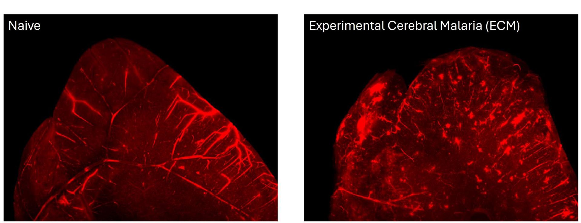
Source publication: Feng G, Li JL, Claser C, Balachander A, Tan Y, Goh CC, Han IKW, Rénia L, Tang BZ, Ng LG, Liu B, Dual modal ultra-bright nanodots with aggregation-induced emission and gadolinium-chelation for vascular integrity and leakage detection, Biomaterials (2017), doi: 10.1016/j.biomaterials.2017.10.031.
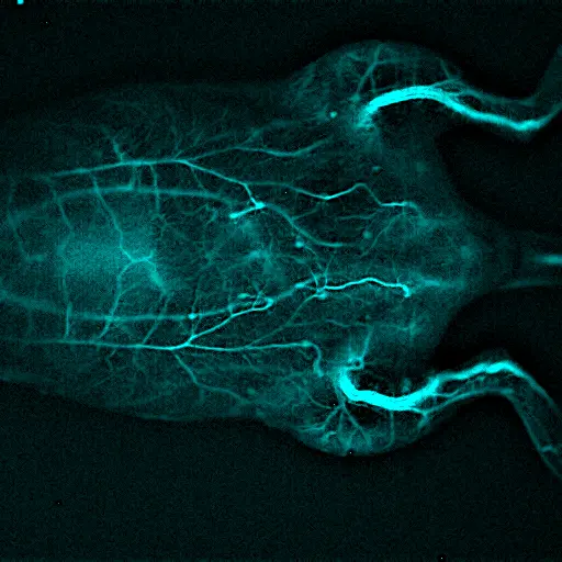
Real-time two-photon blood vasculature imaging in live mice
Source publication: Ding D, Goh CC, Feng G, Zhao Z, Liu J, Liu R, Tomczak N, Geng J, Tang BZ, Ng LG, Liu B. Ultrabright organic dots with aggregation-induced emission characteristics for real-time two-photon intravital vasculature imaging. Adv Mater. 2013 Nov 13;25(42):6083-8. doi: 10.1002/adma.201301938. Epub 2013 Aug 19. PMID: 24038281.
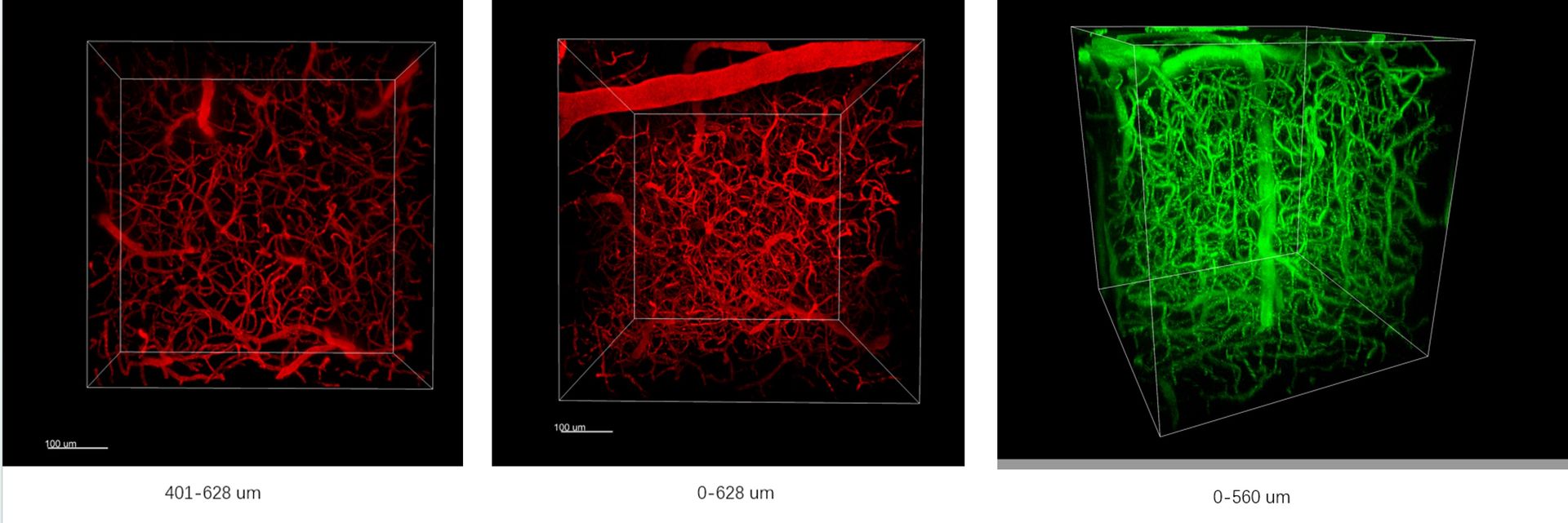
3D reconstruction of vascular systems with two-photon fluorescence imaging
Establishing human models of cerebral cavernous malformations (CCM) deficient 3D vasculature
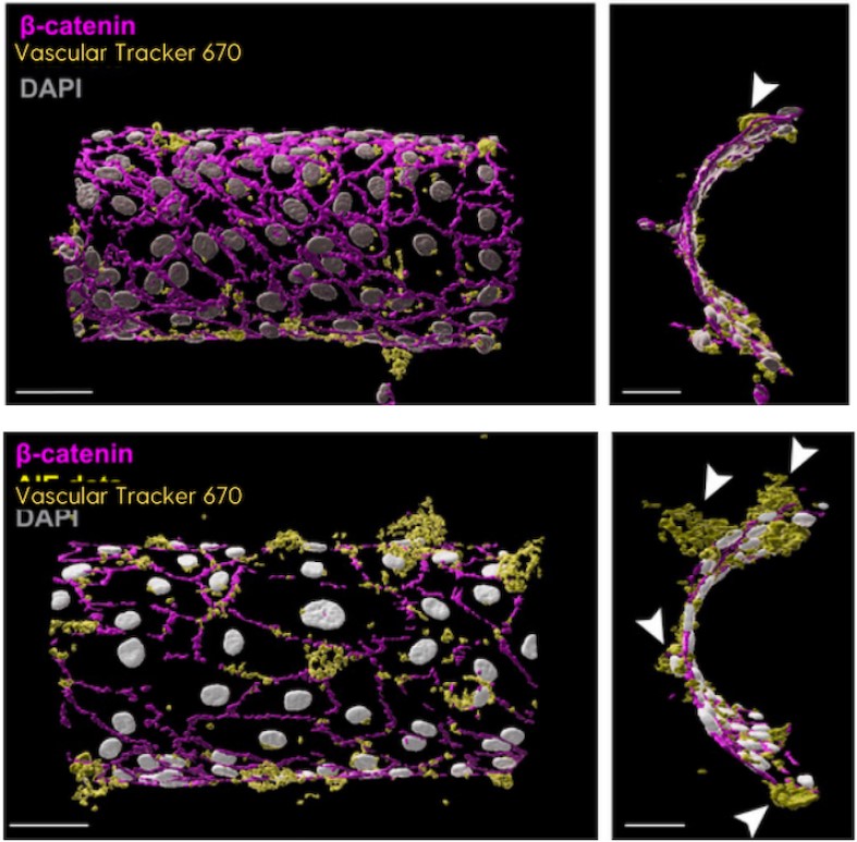
Credits: Dr. Anne Lagendijk & Elysse Moris
Ex vivo organ fluorescence imaging using Vascular Tracker 670
Credits: Kratoscope, Kaer Labs. Courtesy N. Chouin & M Cherel, CRCI2NA Nantes
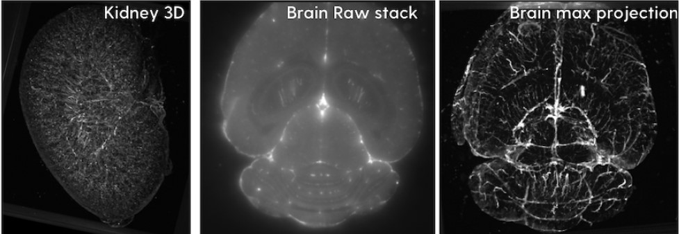
Whole mice vasculature imaging with Vascular Tracker 1010
Credits: Xavier Le Guével & Dr. Véronique Josserand
Other products you may like
Stampwell - Zebrafish
Use Stampwell to facilitate imaging of labelled vasculature in multiple zebrafishes at the same time by immobilizing and aligning them perfectly.
Actiflash
Discover our photoinducible protein activator to precisely control protein activity down to the single-cell resolution in animal models.