GPC4-Nanobody:
a stem cell differentiation booster
Single-domain antagonizing antibody blocking Glypican-4 protein activity and promoting stem cell entry into differentiated lineages
GPC4-Nanobody is a single-domain antagonizing antibody (sdAb) that recognizes and binds a conformational epitope of the native human Glypican-4 (hGPC4) with high affinity and specificity. When added to the culture medium of live cells, GPC4-Nanobody blocks hGPC4 biological functions.
GPC4-Nanobody can be used as a differentiation booster: by maintaining reduced hGPC4 levels throughout differentiation, GPC4-Nanobody addition in the differentiation medium leads to more efficient differentiated lineage entry in response to triggering factors.
A differentiation booster
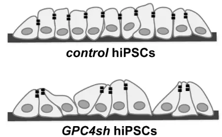
GPC4-Nanobody arises from the groundbreaking discovery that constant reduction in hGPC4 levels in differentiating hiPSCs makes them more responsive to differentiation signals (cf Publications section). Continuous exposure of hiPSC to GPC4-Nanobody allows them to acquire a unique biologial state characterized by:
- Maintenance of pluripotency in undifferentiated conditions (absence of differentiation factors)
- Enhanced differentiation yields upon differentiation exposure
- Reduced tumorigenicity in xenografts
Operating principle of GPC4-nanobody
Increase differenciation
So far, GPC4-Nanobody has been shown to increase differentiation yield of hiPSC into endoderm progenitors and dopaminergic neurons.
Detect hGPC4
GPC4-Nanobody can be used to detect and quantify hGPC4 levels (native or recombinant) with high specificity in immunostaining, flow cytometry or immunoprecipitation assays performed unfer non-denaturing conditions.
This nanobody binds to hGPC4 only in its native conformation: it cannot be used for hGPC4 detection in Western Blots or other assays performed under denaturing conditions.
Block hGPC4
GPC4-Nanobody binds hGPC4 with high affinity and blocks its biological functions: beyond hGPC4 detection, it can also be used to specifically inhibit its activity temporally or permanently in live cells. A simple way to unravel hGPC4 specific roles in various biological contexts without inducing any genetic modifications in mechanistic studies or therapeutic/diagnostic tool development!
Increase your stem cells differentiation yield with GPC4
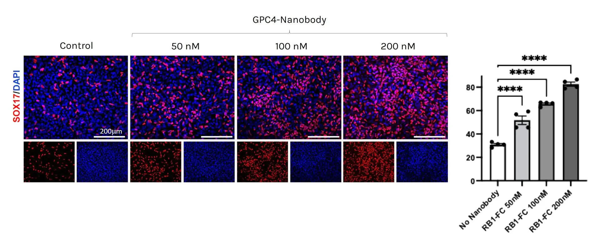
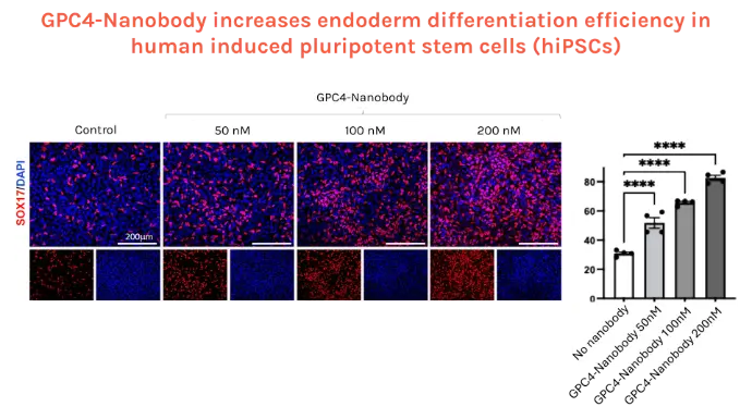
GPC4-Nanobody increases endoderm differentiation efficiency in human induced pluripotent stem cells (hiPSCs)
Left: Immunofluorescence analysis of SOX17 endodermal marker (red) and DAPI (blue) positive cells in hiPSC cultured for 2 days with endodermal differentiation factors alone (Control) or added with GPC4-Nanobody at increasing concentrations. Right: Quantification of the percentage of SOX17+ progenitors over DAPI+ (n = 4 biological replicates).
Credits: Rosanna Dono (Institut du Biologie du Développement de Marseille IBDM, Aix-Marseille Université, France)
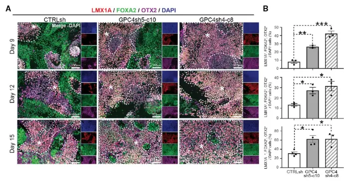
Downregulation of GPC4 in hiPSCs promotes efficient generation of ventral midbrain dopamine neuron progenitors in vitro
A. Immunofluorescence analysis of LMX1A (red), FOXA2 (green), OTX2 (purple), and DAPI (blue) positive cells in CTRLsh, GPC4sh5-c10, and GPC4sh4-c8 cultures at the indicated time points of VMDA differentiation. LMX1A+, FOXA2+, OTX2+ cells, which correspond to bona fide VMDA progenitors, are highlighted by dashed areas with a star. B. Quantification of the percentage of LMX1A+, FOXA2+, OTX2+ VMDA progenitors over DAPI+ (VMDA progenitors at d9: CTRLsh: 8.1 ± 1.8%; GPC4sh5-c10: 26.5 ± 1.6%; GPC4sh4-c8: 42.3 ± 3.1%; at d12: CTRLsh: 13.3 ± 1.2%; GPC4sh5-c10: 27.1 ± 3.1%; GPC4sh4-c8: 31.4 ± 4.6%; at d15: CRTLsh: 31.4 ± 3.1%; GPC4sh5-c10: 62.7 ± 7.6%; GPC4sh4-c8: 62.7 ± 9.0%). Data are presented as mean ± SEM (n = 2-3 biological replicates). One-way ANOVA: *P < .05, **P < .01, ***P < .001.
Source publication: Corti S. et al, 2020
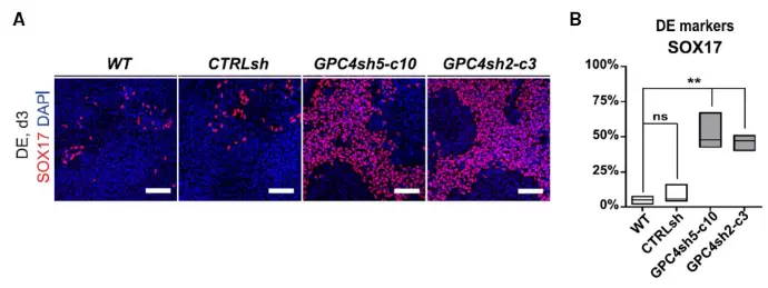
Downregulation of GPC4 in hiPSCs enhances differentiation potential into definitive endoderm
A. Immunofluorescence analysis of SOX17 (red, DE) onWT, CTRLsh, GPC4sh5-c10 and GPC4sh2-c3 029 hiPSCs differentiated for 3 days into DE, n = 3. Scale bar: 100 μm. B. Percentages of SOX17 positive cells in WT, CTRLsh, GPC4sh5-c10 and GPC4sh2-c3 029 hiPSCs were quantified from staining shown in A. Box plots represent the median with min and max values, n = 3. Statistical analysis: two-way ANOVA, followed by Dunnett’s multiple comparison test. P-values: (***) <0.001, (**) <0.01, (*) <0.05, ns not significant.
Source publication: Legier et al, 2023
The use of GPC4 as a stem cell differentiation booster is supported by several publications:
Remi Bonjean, Rossana Cuciniello, Brigitte Kerfelec, Patrick Chames, Rosanna Dono, Functional targeting of Glypican-4 by a conformation-specific single-domain antibody,Biomaterials, 2025, 123864, ISSN 0142-9612, https://doi.org/10.1016/j.biomaterials.2025.123864.
Legier, T., Rattier, D., Llewellyn, J. et al. Epithelial disruption drives mesendoderm differentiation in human pluripotent stem cells by enabling TGF-β protein sensing. Nat Commun 14, 349 (2023). https://doi.org/10.1038/s41467-023-35965-8
Corti S, Bonjean R, Legier T, Rattier D, Melon C, Salin P, Toso EA, Kyba M, Kerkerian-Le Goff L, Maina F, Dono R. Enhanced differentiation of human induced pluripotent stem cells toward the midbrain dopaminergic neuron lineage through GLYPICAN-4 downregulation. Stem Cells Transl Med. 2021 May;10(5):725-742. doi: 10.1002/sctm.20-0177. Epub 2021 Feb 2. PMID: 33528918; PMCID: PMC8046045.
Publication highlighted on the IBDM Institute's website: https://www.ibdm.univ-amu.fr/a-new-target-to-control-the-differentiation-and-tumorigenic-potentials-of-human-induced-pluripotent-stem-cells/
GPC4-Nanobody is a patented technology (International Publication Number: WO 2022/079270 A1)
You want to know more about this stem cell differentiation booster?
Let's discuss your experiments!