WRAP peptide is a 15-amino acid-long transfecting agent that can WRAP'n ROLL around nucleic acids to form nanoparticles. Those nanoparticles can easily penetrate the cells and deliver their precious cargoes such as plasmids or siRNA
Authors : Prisca Boisguerin - Eric Vives - Gudrun Aldrian - Sebastien Deshayes - Karidia Konate
#peptide #nanoparticle #transfection # RNAinterferenceA few years ago... How I met WRAP (and its publication)
When I was an intern within the public/private joint unit Sys2Diag I met with Gudrun, a peptide chemist with plenty of promising peptide-based projects. Among them were the WRAP peptide series: short peptides (less than 16 amino acids, mostly made of leucine, tryptophan, and arginine residues) with an impressive potential as transfection reagents.
Peptide-Based Nanoparticles to Rapidly and Efficiently “Wrap ’n Roll” siRNA into Cells Bioconjugate Chem. 2019, 30, 592−603
Deciphering the internalization mechanism of WRAP:siRNA nanoparticles Biochimica et Biophysica Acta (BBA) - Biomembranes 2020 Vol 1862, Issue 6
For a quick overview, allow me now to introduce you to those tiny peptidic vectors!

First, why are transfection peptides important?
When I study a research tool I am always wondering: what challenge is awaking these scientists every morning? What unmet need drives them to innovate?
In the case of WRAPs, the challenge is: breaking into the cell unnoticed!
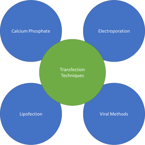
Entering the cell and delivering cargo is a central aspect in plenty of biotechnologies, say for example RNA interference, Plasmid Transfection, Crisp-Cas9 gene editing, the COVID-19 vaccine (!). A few techniques exist to achieve that (see on the left).
Ideally, you want your entry method to be cheap, efficient, and minimally disruptive for the cells. Easier said than done...
Calcium phosphate and electroporation are simple methods, yet can only be applied in vitro. Lipofectamine and viral methods are compatible with in vivo models but are trickier to produce and handle.
Being composed of natural amino acids, peptidic transfecting agents are i) stable in solution ii) able to transfect many cell types with nearly no toxicity iii) open for chemical modification to increase specificity or stability in vivo.
All this added-up makes them very interesting vector molecules.
How do they work?
WRAP peptides work in a 3-step process: 1. Assembly into nanoparticles; 2. Internalization and 3. Delivery of the siRNA/plasmid
1. Nanoparticle Assembly
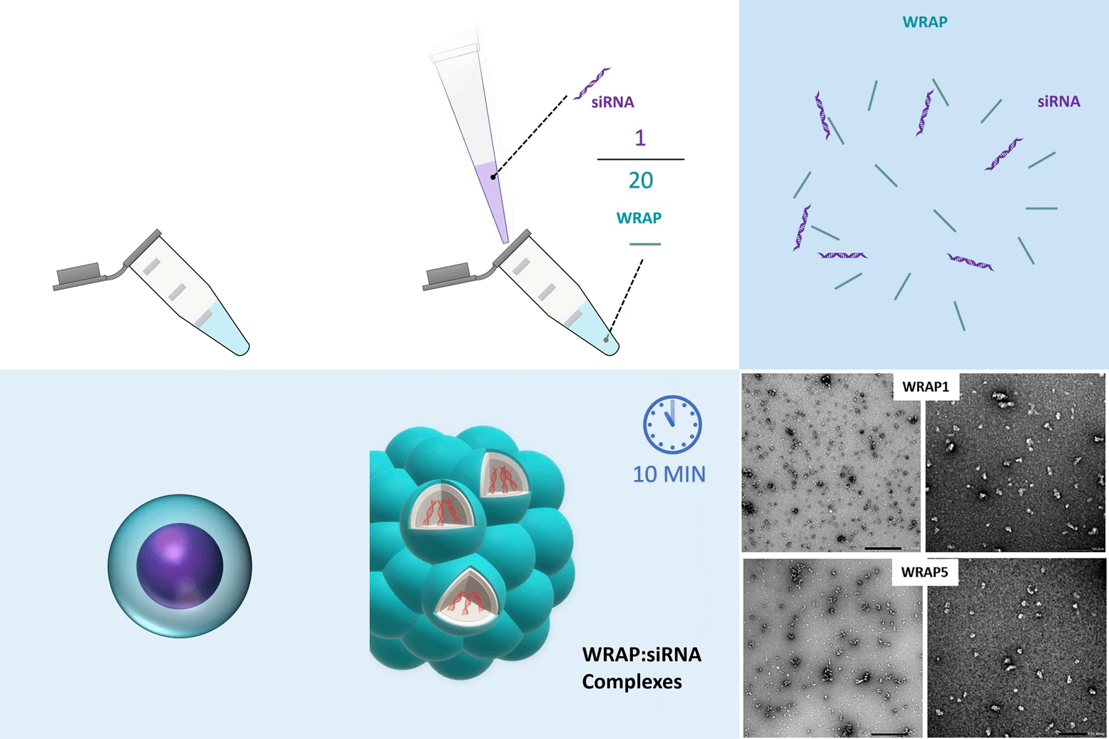
Thanks to their structure and amino acid composition, WRAP peptides bind the nucleic acids in a non-covalent fashion and form peptide-based nanoparticles. This process was extensively studied in their first paper (Bioconjugate Chem. 2019).
Briefly, with the adequate nucleic acid / WRAp ratio, the molecules will self-assemble into small complexes (100 nm) with siRNA at the core and peptide in the shell. Those WRAP:siRNA complexes formed globular nanoparticles as observed by Transmission Electron Microscopy (TEM).


2. Internalization
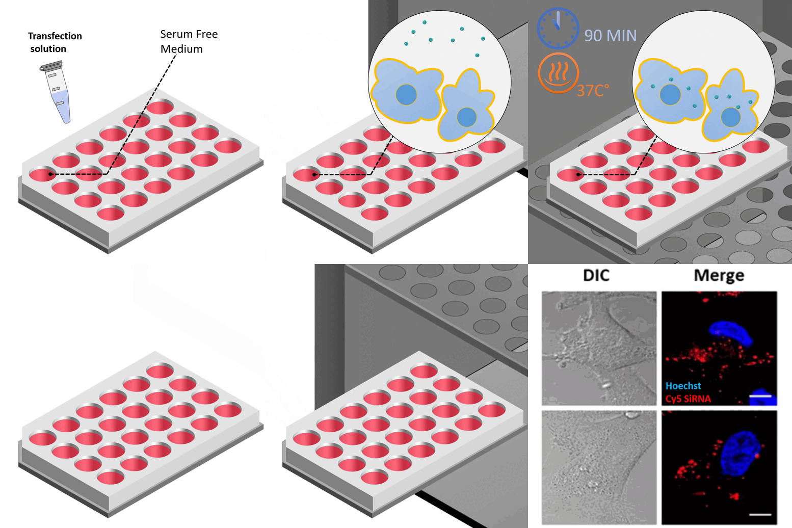
When added to cells, the nanoparticles enter the cell by direct translocation. This process was extensively studied in the second paper (BBA Biomembranes 2020).
The internalization can be visualized using epifluorescence microscopy. Here, a fluorescent siRNA (red) highlights the internalization of the nanoparticle within the cell (nucleus in blue).
3. Gene Knock Down with minimal cell disruption
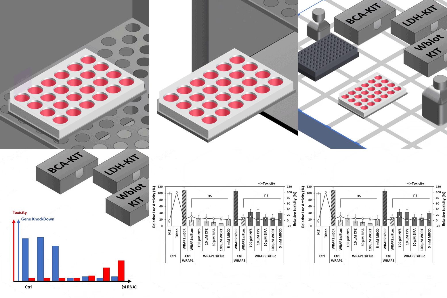
Once inside the cell, the siRNA is released and quickly silences the targeted gene expression. The more WRAP:siRNA complexes the stronger the knockdown, however, as for any vector molecule, it is crucial to perform serial dilution experiments to find the optimal concentration as seen in the example animation on the right.
In a typical experiment, after determination of proteins concentration (BCA assay) and cell viability (LDH kit), a Western Blot reveals the efficient knockdown of the targeted gene.
Conveniently, WRAPs are minimally disruptive to the cells: In U87 cells Luciferase silencing assays, WRAPs nanoparticles displayed energy- and endocytosis-independent internalization mechanism while LDH assay revealed no effect on cell viability.
.gif)
WRAPping up my post
MORE LIFE SCIENCE TOOLS TO FOLLOW
You want the latest on life science tools to follow, test and more?
