
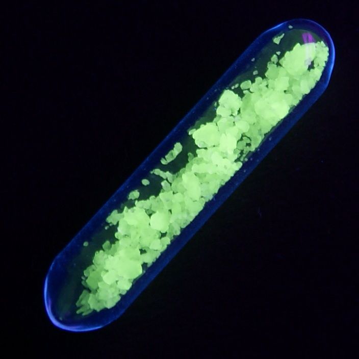
Scientists are coupling terbium nanoparticles with organic ligands that enhance the fluorescence and allow biomolecule labelling. They obtain fluorescent particles that are brighter than quantum dots while also having room for functionnalization. These next generation probes may find use both for in-vitro detection and fluorescence Imaging.
Authors: Dr. Cyrille Charpentier, Dr. Vjona Cifliku, Dr. Joan Goetz, Dr. Aline Nonat, Dr. Clémence Cheignon, Dr. Marcelina Cardoso Dos Santos, Dr. Laura Francés‐Soriano,Prof. Dr. Ka‐Leung Wong, Dr. Loïc J. Charbonnière, Prof. Dr. Niko Hildebrandt
Why using terbium as a fluorescent probe?
For those working with fluorescent probes on biological objects, terbium fluorescent properties provide an answer to a common question:
How do I get rid of the noise coming from my sample?
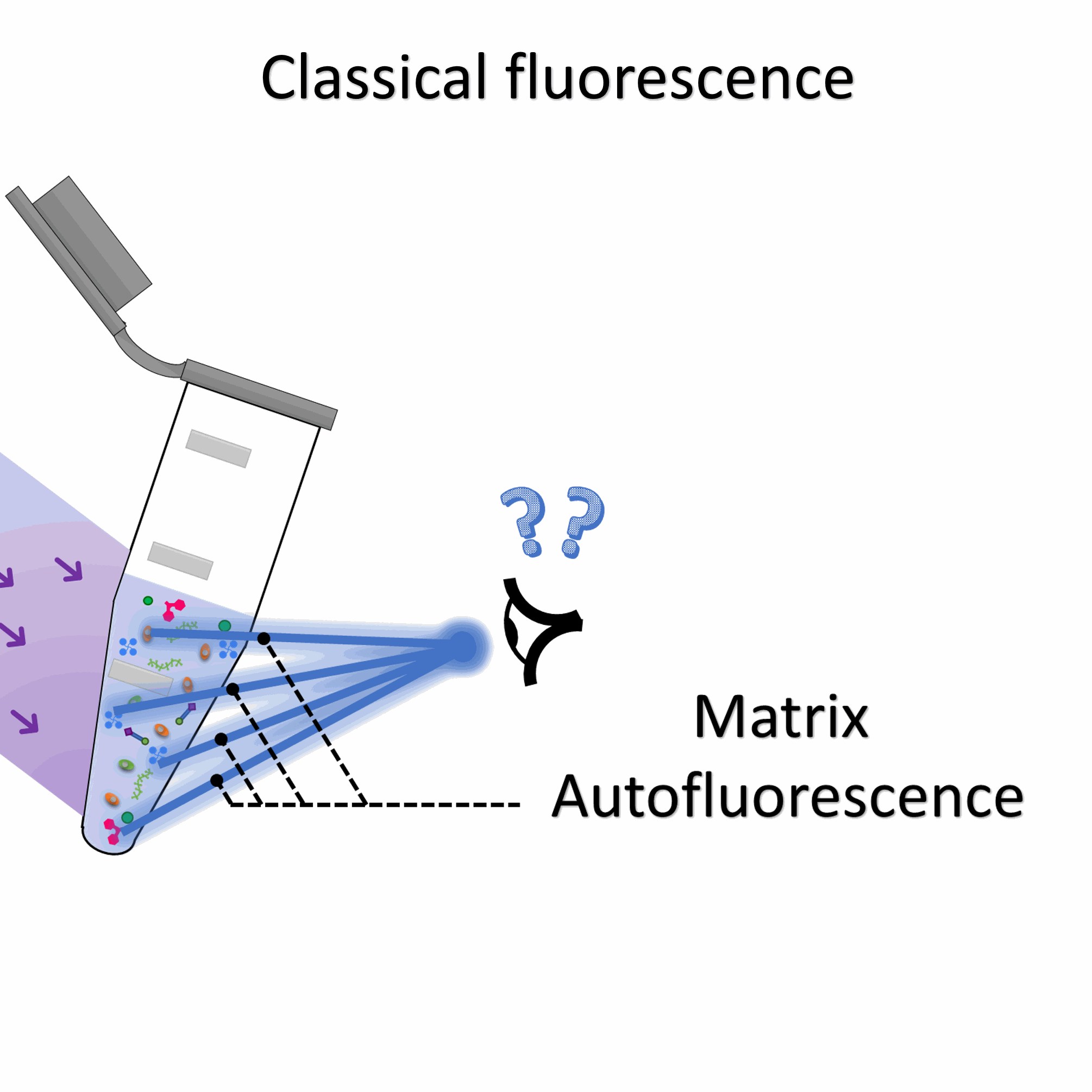
Biology generates background noise: autofluorescence
Biological samples are a mess: the diversity of compounds that can be found inside a cell is ludicrous.Thus, putting a fluorophore inside a biological samples and expecting the rest of the molecules to be stricly non fluorescent is a forlorn hope.
Most of the time, biological matrices display autofluorescence which decreases signal to noise ratio.
Image credit: Aurélien Pasturel 2021
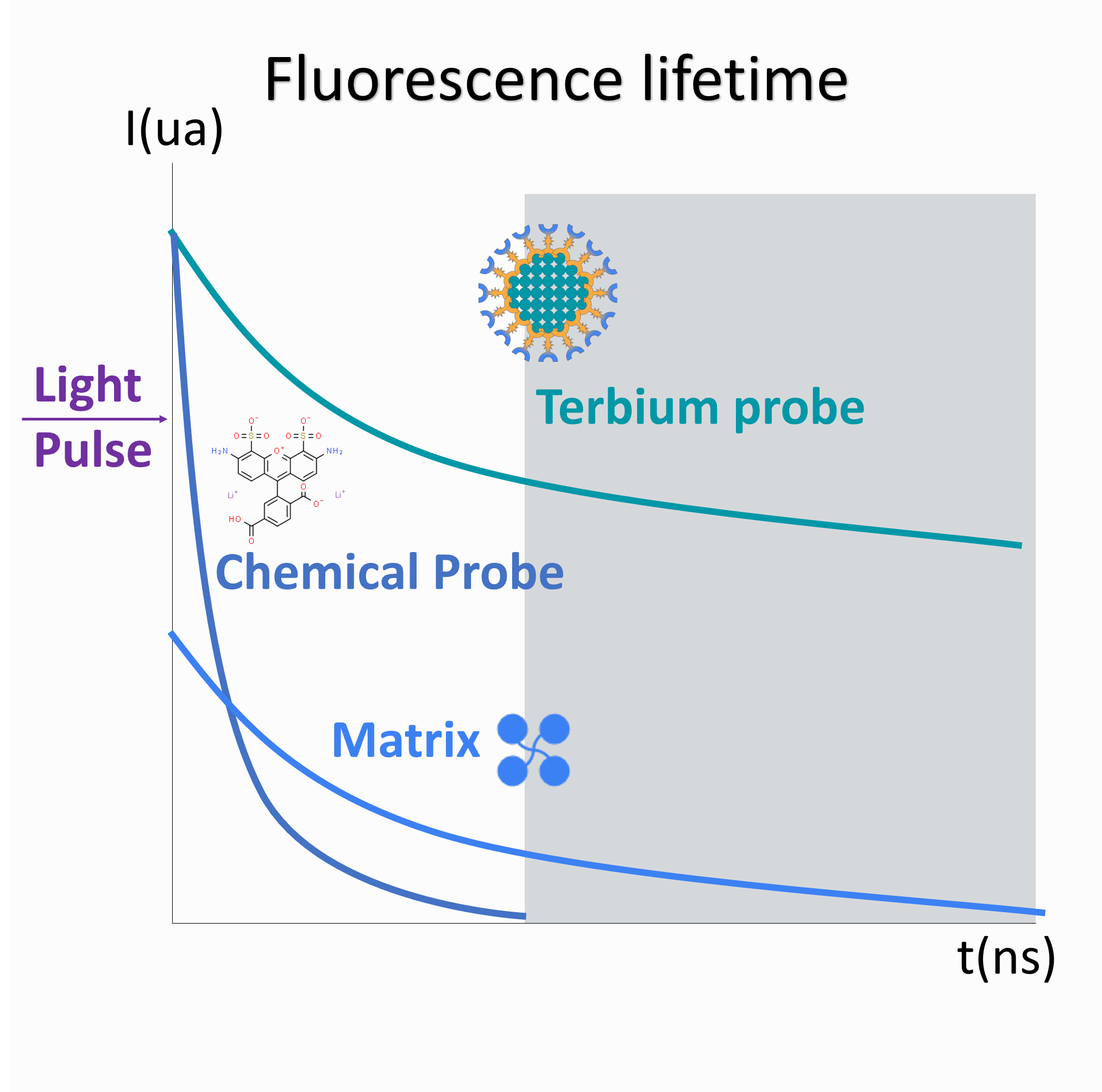
Terbium fluorescence can outlast that of the matrix...
Fluorescence lifetime describes the duration of fluorescent emissions upon excitation with a light pulse.
For most chemical probes, the fluorescence intensity decays exponentially over short (10ns) timeframes.
Interestingly, terbium has a much longer (ms) lifetime, many times superior to chemical probes and matrix autofluorescent compounds.
Image credit: Aurélien Pasturel 2021
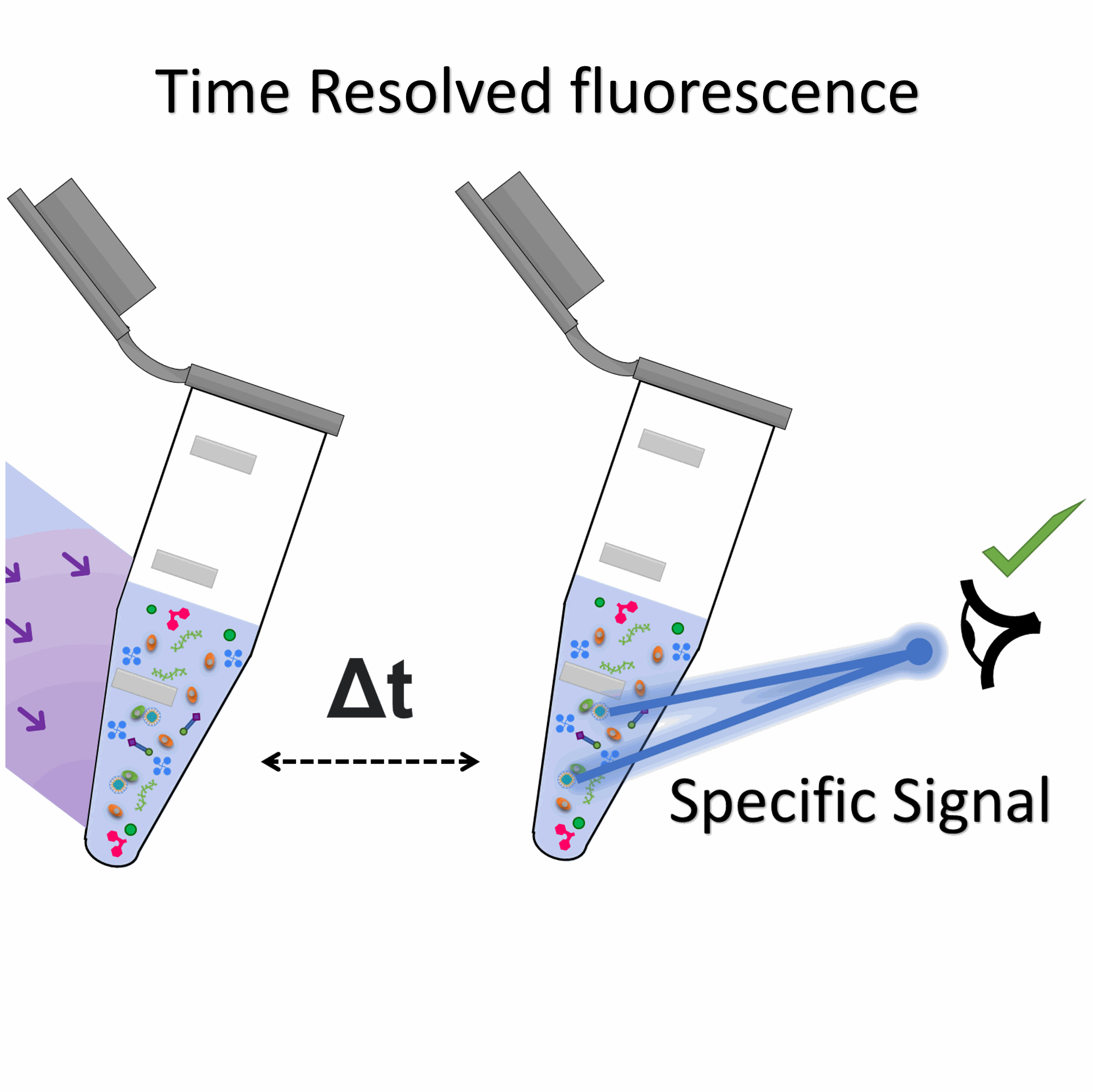
... allowing a delay between excitation and emission
In time-resolved fluorescence experiments: laser excitation and fluorescence recording are delayed.
This delay is sufficient for background fluorescence to disappear while terbium emission is still strong.
This methodology ensures a much higher signal to noise ratio.
Image credit: Aurélien Pasturel 2021
How did Rare Earth Materials become a thing in fluorescence? A brief history
Lanthanides aren't highly fluorescent
on their own
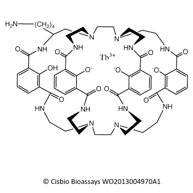
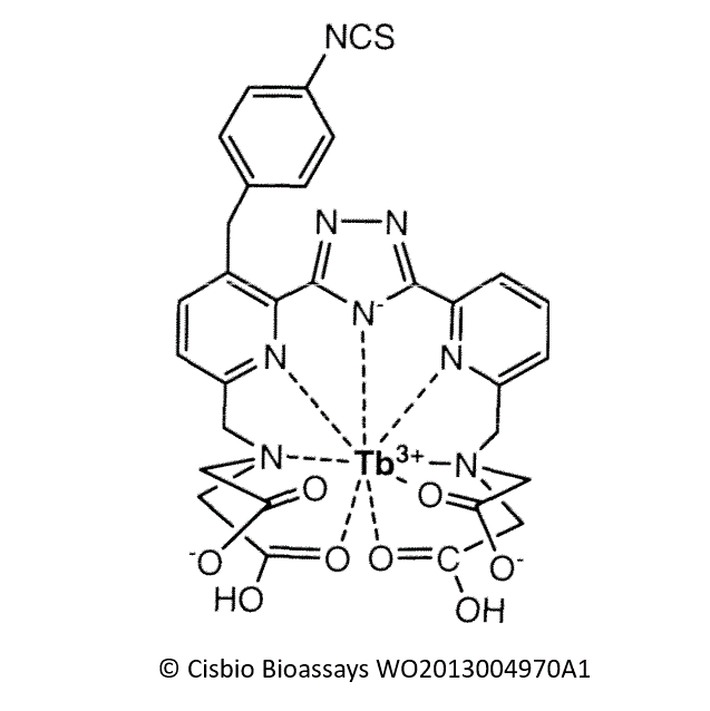
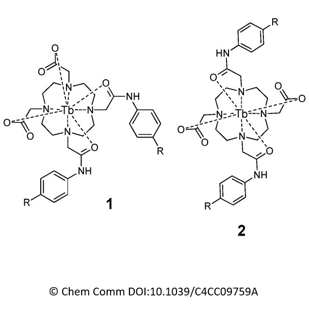
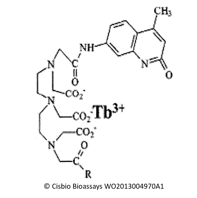
, to harness their fluorescent potential one needs to encapsulate them in molecular cages: cryptates.
Nonetheless, coupling a rare earth material to anything remotely organic is no trivial thing.
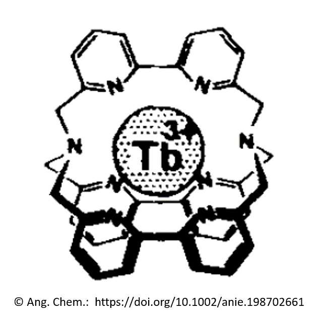
Pioneering works date back from 1987, when Beatrice Alpha, Jean Marie Lehn and Gerard Mathis successfully used cryptate ligands to "cage" single terbium atoms and studied their fluorescent properties.
Since then, a flurry (see below for a non exhaustive list) of a molecules were developed to encapsulate terbium (and other rare earth materials) leading to many publications and patents.
And then, researchers started playing with terbium nanoparticules...
How do they work?
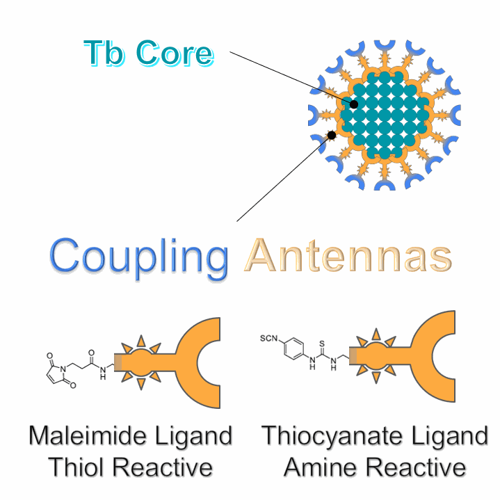
The never-ending need for better probes pushed researchers towards caging whole nanoparticles instead of single lanthanide atoms. This led to the developpement of surface-attached antenna ligands.
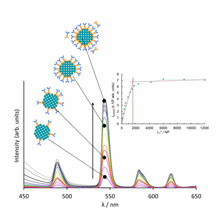
The nanoparticles are coated with fluorescence-enhancing antennas
Much like the cryptate complexes, the terbium nanoparticles must be functionnalized with organic chromophores to dope their fluorescence. These molecules are called "surface-attached antenna ligands".
On the graph above, one can see a titration curve. Upon excitation with a 337 nm laser, terbium fluorescence (note the distinct 4-band spectral signature) is increased by the addition of ligands until it reaches a plateau (around 1700 antennas) signaling a fully coated nanoparticle
Image credit: Aurélien Pasturel 2021 and
Chemistry - A European Journal https://doi.org/10.1002/chem.202002007
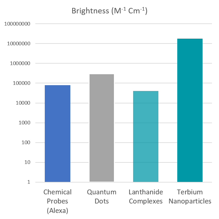
The antennas provide high brightness ...
Single-atom lanthanides complexes are not highly fluorescent and, in fact, display lower fluorescence than standard probes.
On the other hand, the antenna-coated terbium nanoparticles are much brighter, even brighter than the well known Quantum Dots.
To quote researchers working on those nanoparticles:
"Such a terbium nano-particle is as bright as about 340 free terbium complexes."
... and acess for biofunctionnalization
The antennas that were developed by Cyrille Charpentier et al. have a dual function.
On top of increasing the fluorescence, they also carry click chemistry moieties: maleimide or thiocyanate.
Those moieties respectively react with the free thiols and amines from the biomolecules allowing the nanoparticles to be further functionnalized.
Image credit; Aurélien Pasturel 2021 and
Chemistry - A European Journal
https://doi.org/10.1002/chem.202002007
What can we do with these?
BIofunctionnalization opens the road for several applications, the first of which being time gated fluorescence imaging and FRET-Based detection. See below:
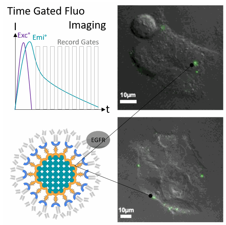
Time-Gated Fluorescence
Time gated fluorescence imaging, is different from traditionnal fluorescence imaging: fluorescence emission is recorded after the laser pulse and over multiple temporal windows (gates) instead of continuously.
This allows researchers to get rid of background noise granted that the fluorescent probe has a sufficient emission lifetime which is totally not an issue with terbium probes.
In the example above, Charpentier et al. coated terbium nanoparticles with EGFR-targeting antibodies to tag the receptor at the surface of the cells.
Interestingly very low UV doses where necessary to obtain a bright signal.
Image credit: Aurélien Pasturel 2021 and Chemistry - A European Journal https://doi.org/10.1002/chem.202002007
FRET-Based detection
Förster Resonance Energy Transfer (FRET) occurs when two fluorophores working as a donor-acceptor pair are nearby.
Upon excitation, the donor will transfer its energy to the acceptor instead of emitting a photon. The acceptor then emits a photon corresponding to its own emission spectra.
As a proof of concept , the researchers coated the nanoparticles with Streptavidin and used a biotynilated A647 to trigger FRET. In the titration experiments, the fluorescence of terbium decreased and while the signal for Alexa Fluor increased as the nanoparticles became fully coated with the biotinylated probe validating the FRET.
Image credit: Aurélien Pasturel 2021 and Chemistry - A European Journal https://doi.org/10.1002/chem.202002007
Closing: Going the distance with fluorescent probes
It is fascinating to see how fluorescent probes have evolved in the last 30 years.
Now researchers have access to probes that are easy to functionnalize, brighter, immune to photobleaching, that have much longer lifetimes, that can switch emission properties, that do not require much photons, that can sense the pH / the viscosity / the oxygen and the lists goes on.
Among those, the terbium nanoparticles check many of the boxes ... promising !
I hope you enjoyed the read and expect more posts on fluorescent probes in the future!
MORE LIFE SCIENCE TOOLS TO FOLLOW
You want the latest on life science tools to follow, test and more?