Who is Gaëlle Recher ?
"I obtained my PhD in 2010 from Rennes University (with F. Tiaho), where I studied xenopus larvae with 2-photon microscopy. I then joined the group of N. Peyriéras (Gif-sur-Yvette) to investigate cell dynamics in live zebrafish embryos, sharpening my skills to babysit embryos to make sure they are happy and comfortable under the scope. I joined the group of M. Zernicka-Goetz (Cambridge, UK), where I learnt techniques related to mammal embryos. There I realized how demanding these bunch of cells are when it deals with microscopy. I met 3D in vitro systems, and moved to Bordeaux in 2015 to work with P. Nassoy and the alginate capsules! Here, as a CNRS researcher, I study these balls of cells, and still develop microscopy approaches. It was obvious to me to muster up the techniques specifically developed by each community and that's how the "Mount(n)" aka Stampwell was born."Turning curiosity into innovation
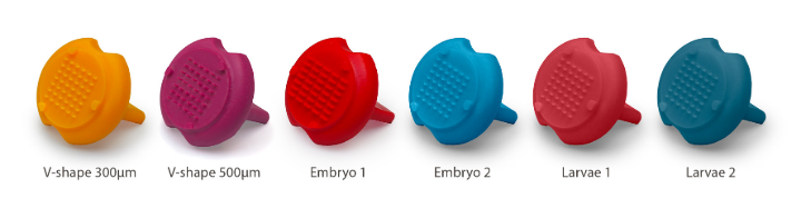
From cell self-organization to tissue biomechanics, she’s not just asking questions, she’s inventing the tools to answer them. Case in point: Stampwell, a tool that simplifies imaging experiments by printing arrays of wells into hydrogels in a matter of seconds and therefore saving researchers countless hours.
Stampwell fun fact
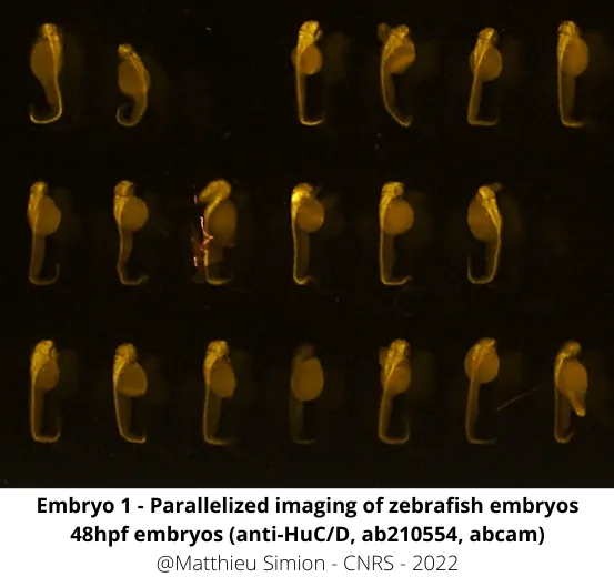
Stampwell was originally designed as a tool to imprint wells for spheroids and organoids. But then researchers asked for a zebrafish version, one that could position zebrafish perfectly for imaging. And that’s how Stampwell-Z was born !
Are you a researcher and want to save time in..
Gaëlle Recher's publications
Andrique, L*, Recher, *, Alessandri, K, Pujol, N, Feyeux, M, Bon, P, Cognet, L, Nassoy, P, and Bikfalvi, A (2019). A model of guided cell self-organization for rapid and spontaneous formation of functional vessels. Science Advances, eaau6562
https://doi.org/10.1126/sciadv.aau6562
Shahbazi M, Scialdone A, Skorupska N, Weberling A, Recher G, Zhu M, Jedrusik A, Devito L, Noli L, Macaulay I, Buecker C, Khalaf Y, Ilic D, Voet T, Marioni J, Zernicka-Goetz M (2017) Pluripotent state transitions coordinate morphogenesis in mouse and human embryos. Nature
https://doi.org/10.1038/nature24675
Alessandri K*, Andrique L*, Feyeux M*, Bikfalvi A, Nassoy P & Recher G† (2017) All-in-one 3D printed microscopy chamber for multidimensional imaging, the UniverSlide. Scientific Reports
https://doi.org/10.1038/srep42378
Joly J-S, Recher G, Brombin A, Ngo K & Hartenstein V (2016) Conveyor-belt neurogenesis: a conserved mode of generating a complex, homotopically patterned visual system in insects and vertebrates. Current Biology
https://doi.org/10.1016/j.cub.2016.08.017
Shahbazi M*, Jedrusik A*, Vuoristo S*, Recher G, Hupalowska A, Campbell A, Fishel S & Zernicka-Goetz M (2016) Self-organization of the human embryo in the absence of maternal tissues. Nature Cell Biology
https://doi.org/10.1038/ncb3347
Faure E*, Savy T*, Rizzi B*, Melani C*, Stasova O*, Fabrèges D*, Spir R, Hammons M, Cunderlik R, Recher G, Lombardot B, Duloquin L, Colin I, .../..., Peyriéras N & Bourgine P (2016) An algorithmic workflow for the automated processing of 3D+time microscopy images of developing organisms and the reconstruction of their cell lineage. Nature Communications
https://doi.org/10.1038/ncomms9674
Recher G*, Jouralet J*, Brombin A, Heuzé A, Mugniery E, Hermel J-M, Desnoulez S, Savy T, Herbomel P, Bourrat F, Peyriéras N, Jamen F & Joly J-S (2013) Zebrafish midbrain slow-amplifying progenitors exhibit high levels of transcripts for nucleotide and ribosome biogenesis. Development
https://doi.org/10.1242/dev.099010
Rouède D, Bellanger J-J, Schaub E, Recher G & Tiaho F (2013) Theoretical and Experimental SHG Angular Intensity Patterns from Healthy and Proteolysed Muscles. Biophysical Journal
https://doi.org/10.1016/j.bpj.2013.02.047
Recher G, Rouède D, Schaub E & Tiaho F (2011) Skeletal muscle sarcomeric SHG patterns photo-conversion by femtosecond infrared laser. Biomedical Optics Express
https://doi.org/10.1364/BOE.2.000374