
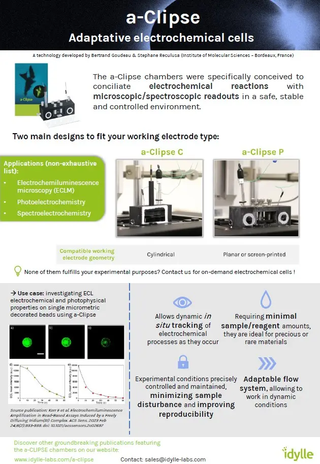
A technology developed by Bertrand Goudeau & Stephane Reculusa
(Institute of Molecular Sciences - Bordeaux, France)
Monitoring electrochemical reactions in situ can prove difficult.
a-Clipse is a set of adaptative chambers, implementable on commonly used microscope and spectrometer instruments, that offer the possibility to conciliate electrochemical reactions and microscopic/spectroscopic readouts in a safe, stable and controlled environment.
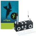
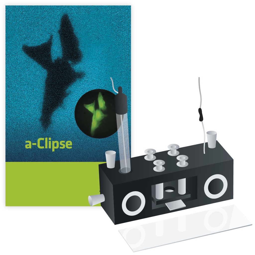
a-Clipse at a glance
The a-Clipse chambers have been specifically designed to make the most of your electrochemical experiments:
in situ monitoring
Allows dynamic in situ tracking of electrochemical processes as they occur
Minimal reagents
Requiring minimal sample/reagent amounts, they are ideal for precious or rare materials
Precise control
Experimental conditions are precisely controlled and maintained in an air-tight container, minimizing sample disturbance and improving reproducibility
Compatible with dynamic assays
Adaptable flow system allowing to work in dynamic conditions
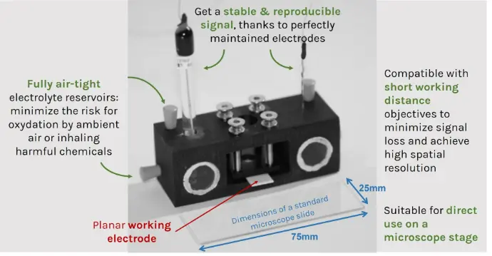
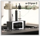
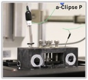
a-Clipse P conceived for inverted microscopes
Ready to take your electrochemical experiments to the next level?
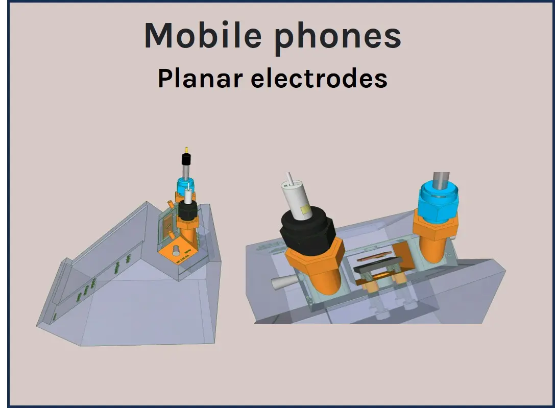
Product specifications
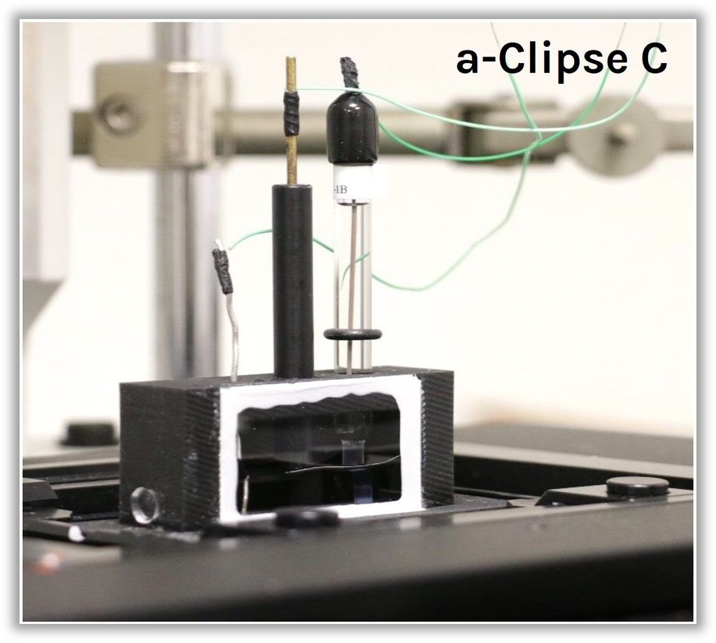
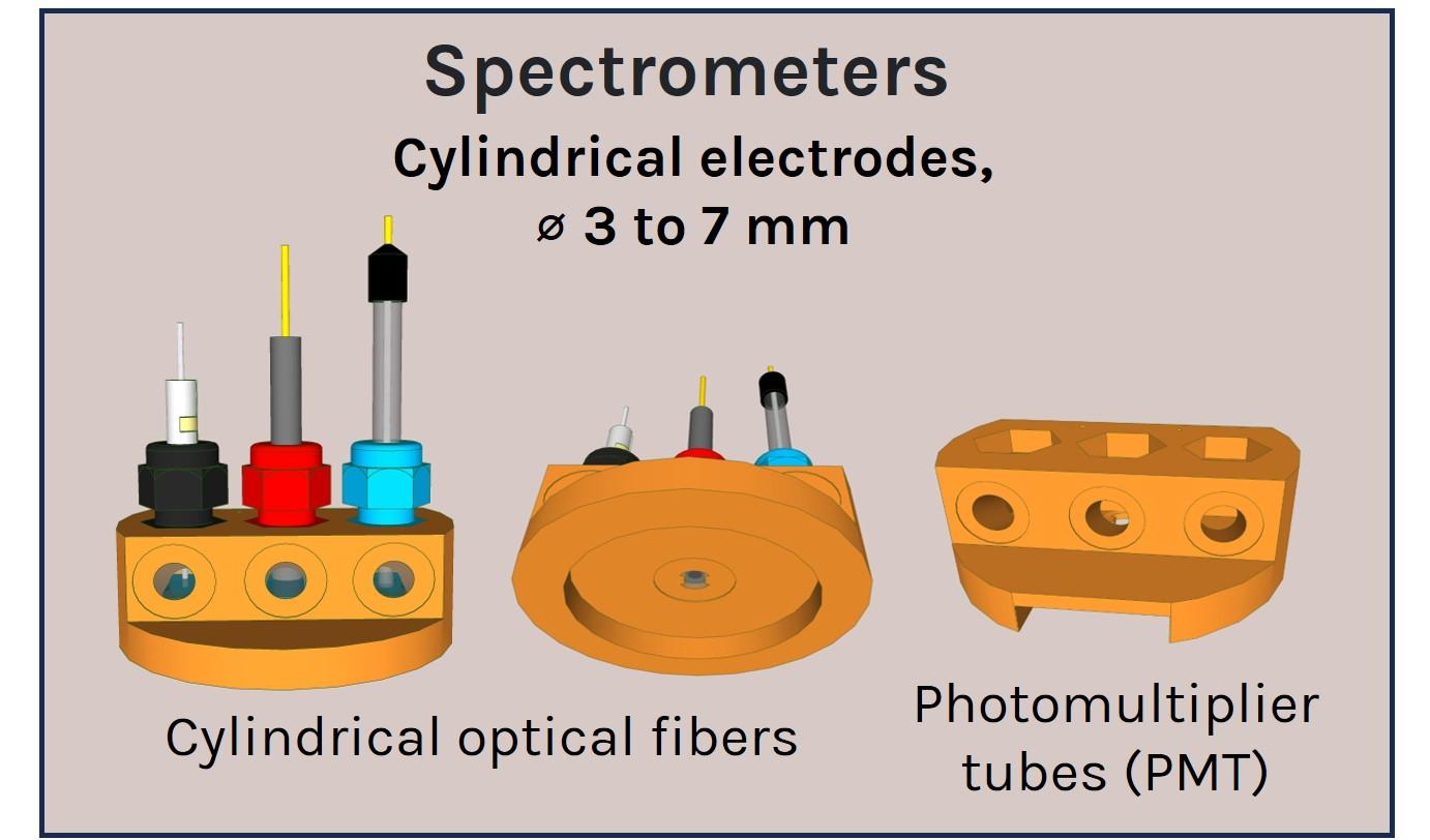
A panel of a-Clipse chambers, compatible with cylindrical and planar working electrode systems, is available to fit a variety of microscope and spectroscope instruments:
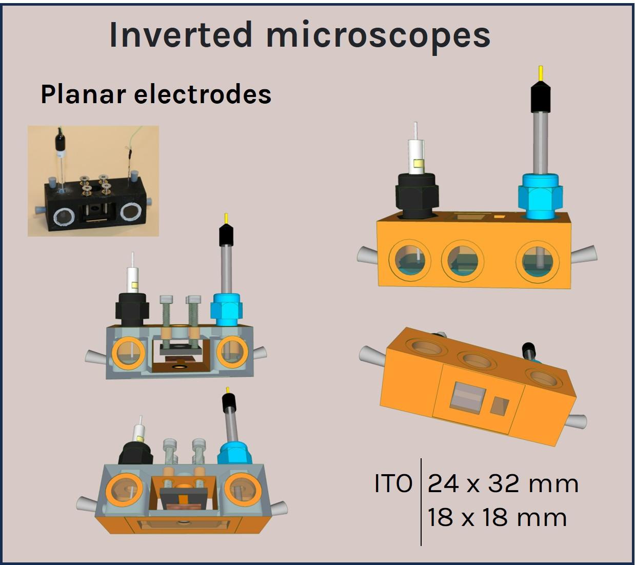
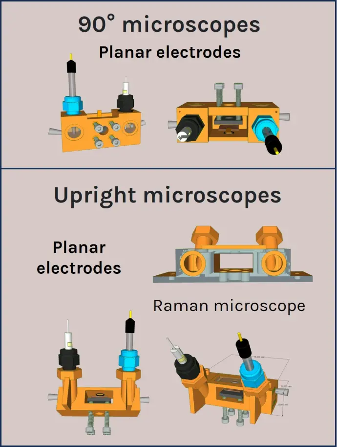
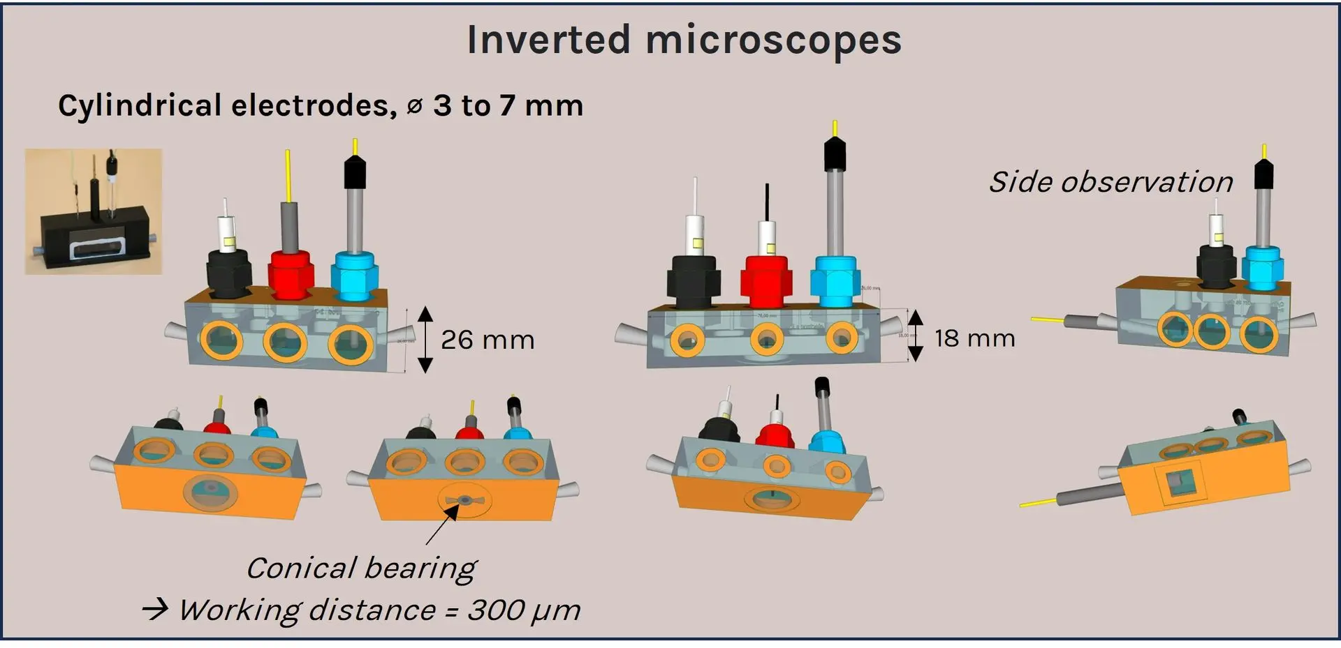
All a-Clipse chambers made for use with microscopes are made with dimensions of a standard microscope slide (25mm x 75mm) holder.
New ! None of them fulfills your experimental purposes? Contact us for on-demand electrochemical cells !
Kit contents
a-Clipse
chambers are provided with all small accessories that are required for their use. Electrodes are not included.
• Electrochemical cell
• Tapered plugs
• LUER connectors
• O-ring (for
a-CLIPSE P chambers only)
• Spare coverslips
Applications
Compatible assays (non-exhaustive list):
- Electrochemiluminescence microscopy (ECLM)
- Photoelectrochemistry
- Spectroelectrochemistry
a-Clipse
chambers can be used to image cells, micro-/nano-objects and electrochemical processes at electrode surfaces. So far,
a-Clipse
chambers have been mostly used in ECLM experiments but can be used for any assay aiming at visualizing electrochemical processes.
Some examples of ultra-sensitive ECLM applications
a-Clipse
chambers contributed to include:
- Imaging single cells & isolated subcellular organelles (i.e. cell membrane proteins & transport, cell-cell contacts, individual mitochondria imaging, etc)
- Development & optimization of (bio)analytical assays (i.e. map the spatial distribution of ECL emission at the surface of single micrometric beads)
- Investigating fundamentals of ECL (i.e. gain deeper insights into the ECL reaction & emission stability mechanisms)
Find out more details and example pictures in our Results section.
Additional resources
> Product overview
> Technical Datasheet
Any other questions? Please contact us.
Infrared Photoinduced Electrochemiluminescence Microscopy of Single Cells
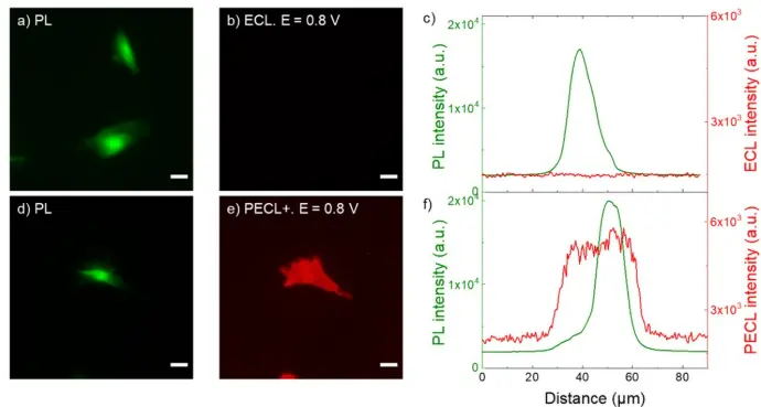
a,d) PL micrographs (green color) of CHO-K1 cells labeled with SA@Ru. b) ECL and e) PECL+ images (red color) of the corresponding labeled cells recorded in ProCell under near-infrared (λexc = 1050 nm) back-illumination at 0.8 V on b) p++-Si/SiOx/Ir and e) n-Si/SiOx/Ir electrodes. c,f) Comparison of the luminescence intensity profiles of the cells in PL, c) ECL and in f) PECL+. The axis along which the profiles were extracted are shown in Figure S5. Green and red are false colors coding the luminescence intensity. Cells were grown on the Ir surfaces, fixed, permeabilized with Triton X-100 and then labeled with SA@Ru. Scale bar: 20 µm. Images obtained with an a-Clipse P chamber for planar electrodes.
Credits: Julie Descamps et al, 2023
Using a-CLIPSE to develop a bimodal electrochemiluminescence imaging approach
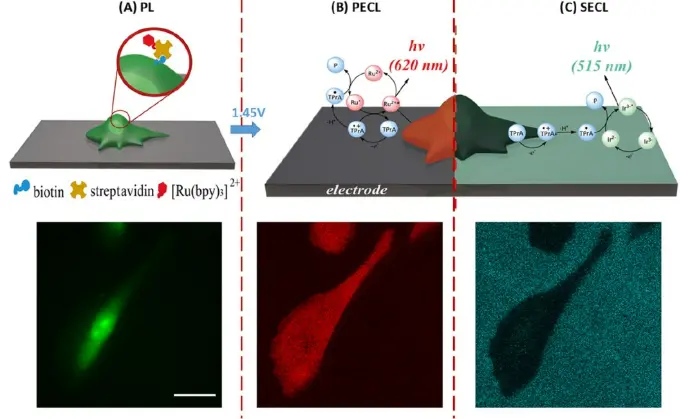
ECLIPSE chambers were used to demonstrate the potential of combining simultaneous positive (PL) and shadow ECL (PECL) configurations on single cells to gain insight into transport properties through permeabilized cell membranes. (Top) Schematic representation of multimodal imaging of a cell immobilized on a glassy carbon electrode (GCE): (A) PL, (B) PECL, and (C) SECL. Mechanisms of co-reactant PECL (heterogeneous route involving mainly dissolved TPrA and SA@Ru label immobilized on the cell) and SECL (homogeneous route involving only dissolved [Ir(sppy)3]3− and TPrA) modes. Ru2+ and Ir3− represent the ECL SA@Ru label and [Ir(sppy)3]3−, respectively. (Bottom) The same single CHO-K1 cell was imaged by (A) PL, (B) PECL, and (C) SECL. Images acquired in a-CLIPSE C chambers.
Credits: Sara Knezevic et al, 2023
Imaging plasma membranes at the single cell level using ECL microscopy
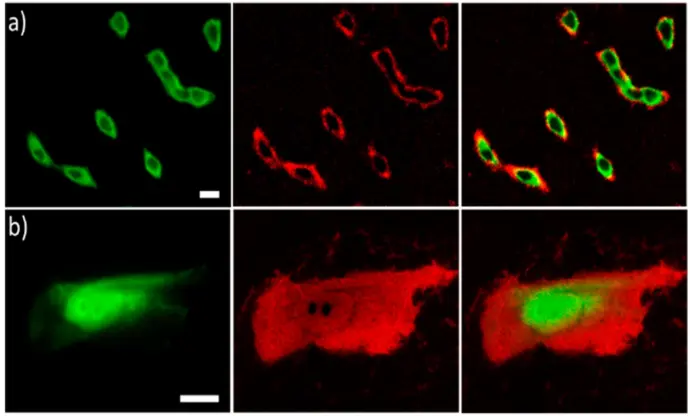
The ECLIPSE chambers were used to specifically image basal membrane details that are not resolved using classic fluorescence microscopy. PL (green), ECL (red) and overlay of both luminescence signals (from left to right) of CHO cells that were (a) labeled with SA@Ru or (b) permeabilized and then labeled with SA@Ru. Cells were grown on GC electrodes. SA@Ru labels were attached to the biotinylated proteins of the cellular membrane. Both PL and ECL images were recorded in the reflection configuration on the same regions of interest in the ECL focal plane. ECL was generated in PBS (pH = 7.4) containing 100 mM TPA by applying 1.4 V. Scale bar: 20 μm.
Images acquired in a-CLIPSE C chambers.
Credits: Silvia Voci et al, 2018
Individual mitochondria imaging in the a-CLIPSE chambers
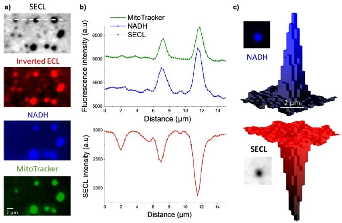
a) Typical SECL and fluorescence images recorded at high magnification of the same ROI showing single mitochondria. b) Luminescence intensity profiles extracted along the dashed line materialized on the top image in (a). c) An example of 3D imaging of a single mitochondrion recorded by NADH fluorescence (top) and SECL (bottom). Images acquired in a-CLIPSE C chambers.
Credits: Yumeng Ma et al, 2021
Using a-CLIPSE to investigate ECL electrochemical and photophysical properties on single micrometric decorated beads
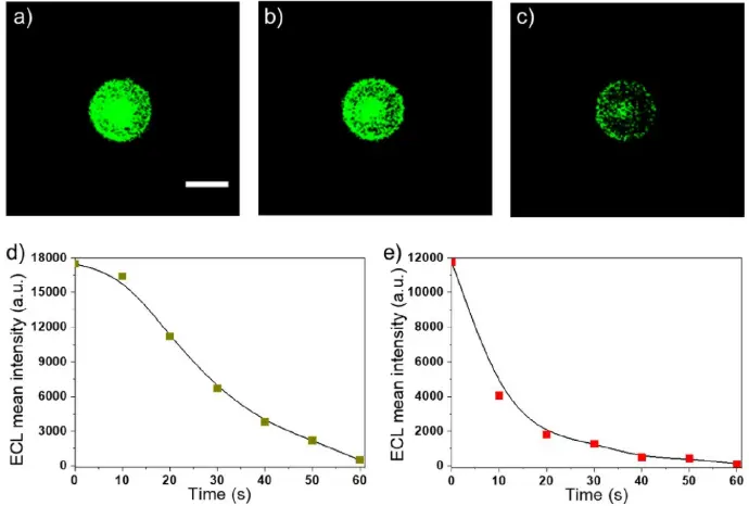
a-c) Sequence of successive ECL images of a single labeled bead recorded in the top-view configuration when imposing a constant potential of 1.1 V. PS 12 μm beads decorated with the ECL label were used. Exposure time: 10 s. Scale bar: 10 μm. Evolution of the ECL intensity with time on d) GC and e) gold electrode when applying a constant potential of 1.1 V. Experiments were performed with a GC or gold working electrode in a PBS solution containing 200 mM TPA (pH 7.4). Experiments have been repeated on more than 30 single beads under each set of conditions. (For interpretation of the references to colour in this figure legend, the reader is referred to the Web version of this article.)
Credits: Priyanka Dutta et al, 2020